| Entry | Database: PDB / ID: 4c13
|
|---|
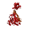
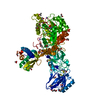
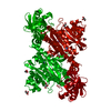
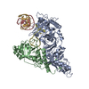
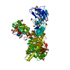
| Title | x-ray crystal structure of Staphylococcus aureus MurE with UDP-MurNAc- Ala-Glu-Lys |
|---|
 Components Components | UDP-N-ACETYLMURAMOYL-L-ALANYL-D-GLUTAMATE--L-LYSINE LIGASE |
|---|
 Keywords Keywords | LIGASE / MURE |
|---|
| Function / homology |  Function and homology information Function and homology information
UDP-N-acetylmuramoyl-L-alanyl-D-glutamate-L-lysine ligase / UDP-N-acetylmuramoyl-L-alanyl-D-glutamate-L-lysine ligase activity / ligase activity / peptidoglycan biosynthetic process / cell wall organization / regulation of cell shape / cell division / magnesium ion binding / ATP binding / cytoplasmSimilarity search - Function UDP-N-acetylmuramoylalanyl-D-glutamate-2,6-diaminopimelate ligase / MurE/MurF, N-terminal / MurE/MurF, N-terminal domain / Mur ligase family, catalytic domain / Udp-n-acetylmuramoylalanyl-d-glutamate--2,6- Diaminopimelate Ligase; Chain: A, domain 1 / Mur ligase, C-terminal domain / Mur-like, catalytic domain / Mur ligase, C-terminal / Mur ligase, C-terminal domain superfamily / Mur ligase, glutamate ligase domain ...UDP-N-acetylmuramoylalanyl-D-glutamate-2,6-diaminopimelate ligase / MurE/MurF, N-terminal / MurE/MurF, N-terminal domain / Mur ligase family, catalytic domain / Udp-n-acetylmuramoylalanyl-d-glutamate--2,6- Diaminopimelate Ligase; Chain: A, domain 1 / Mur ligase, C-terminal domain / Mur-like, catalytic domain / Mur ligase, C-terminal / Mur ligase, C-terminal domain superfamily / Mur ligase, glutamate ligase domain / Mur ligase, central / Mur-like, catalytic domain superfamily / Mur ligase middle domain / UDP-N-acetylmuramoyl-L-alanine:D-glutamate ligase / Protein-Tyrosine Phosphatase; Chain A / Alpha-Beta Complex / 3-Layer(aba) Sandwich / Alpha BetaSimilarity search - Domain/homology Uridine 5'Diphospho N-acetyl muramoyl-L-Alanyl-D-Glutamyl-L-Lysine / : / PHOSPHATE ION / Chem-UML / : / UDP-N-acetylmuramoyl-L-alanyl-D-glutamate--L-lysine ligaseSimilarity search - Component |
|---|
| Biological species |   STAPHYLOCOCCUS AUREUS (bacteria) STAPHYLOCOCCUS AUREUS (bacteria) |
|---|
| Method |  X-RAY DIFFRACTION / MOLECULAR REPLACEMENT / Resolution: 1.9 Å X-RAY DIFFRACTION / MOLECULAR REPLACEMENT / Resolution: 1.9 Å |
|---|
 Authors Authors | Ruane, K.M. / Roper, D.I. / Fulop, V. / Barreteau, H. / Boniface, A. / Dementin, S. / Blanot, D. / Mengin-Lecreulx, D. / Gobec, S. / Dessen, A. ...Ruane, K.M. / Roper, D.I. / Fulop, V. / Barreteau, H. / Boniface, A. / Dementin, S. / Blanot, D. / Mengin-Lecreulx, D. / Gobec, S. / Dessen, A. / Dowson, C.G. / Lloyd, A.J. |
|---|
 Citation Citation | Journal: Nat.Chem.Biol. / Year: 2021Title: Discovery of a first-in-class CDK2 selective degrader for AML differentiation therapy. Authors: Wang, L. / Shao, X. / Zhong, T. / Wu, Y. / Xu, A. / Sun, X. / Gao, H. / Liu, Y. / Lan, T. / Tong, Y. / Tao, X. / Du, W. / Wang, W. / Chen, Y. / Li, T. / Meng, X. / Deng, H. / Yang, B. / He, ...Authors: Wang, L. / Shao, X. / Zhong, T. / Wu, Y. / Xu, A. / Sun, X. / Gao, H. / Liu, Y. / Lan, T. / Tong, Y. / Tao, X. / Du, W. / Wang, W. / Chen, Y. / Li, T. / Meng, X. / Deng, H. / Yang, B. / He, Q. / Ying, M. / Rao, Y. #1: Journal: J.Biol.Chem. / Year: 2013Title: Specificity Determinants for Lysine Incorporation in Staphylococcus Aureus Peptidoglycan as Revealed by the Structure of a Mure Enzyme Ternary Complex. Authors: Ruane, K.M. / Lloyd, A.J. / Fulop, V. / Dowson, C.G. / Barreteau, H. / Boniface, A. / Dementin, S. / Blanot, D. / Mengin-Lecreulx, D. / Gobec, S. / Dessen, A. / Roper, D.I. |
|---|
| History | | Deposition | Aug 9, 2013 | Deposition site: PDBE / Processing site: PDBE |
|---|
| Revision 1.0 | Oct 2, 2013 | Provider: repository / Type: Initial release |
|---|
| Revision 1.1 | Oct 9, 2013 | Group: Database references |
|---|
| Revision 1.2 | Nov 27, 2013 | Group: Database references |
|---|
| Revision 2.0 | Jun 28, 2017 | Group: Advisory / Atomic model / Data collection
Category: atom_site / diffrn_source / pdbx_unobs_or_zero_occ_atoms
Item: _atom_site.B_iso_or_equiv / _atom_site.Cartn_x ..._atom_site.B_iso_or_equiv / _atom_site.Cartn_x / _atom_site.Cartn_y / _atom_site.Cartn_z / _atom_site.auth_atom_id / _atom_site.label_atom_id / _atom_site.type_symbol / _diffrn_source.type |
|---|
| Revision 2.1 | Sep 13, 2017 | Group: Data collection / Category: diffrn_source
Item: _diffrn_source.pdbx_wavelength_list / _diffrn_source.source |
|---|
| Revision 2.2 | Jun 20, 2018 | Group: Data collection / Structure summary / Category: pdbx_molecule_features |
|---|
| Revision 2.3 | Mar 17, 2021 | Group: Database references / Derived calculations / Other
Category: citation / citation_author ...citation / citation_author / pdbx_database_status / pdbx_struct_conn_angle / struct_conn / struct_site
Item: _pdbx_database_status.status_code_sf / _pdbx_struct_conn_angle.ptnr1_auth_comp_id ..._pdbx_database_status.status_code_sf / _pdbx_struct_conn_angle.ptnr1_auth_comp_id / _pdbx_struct_conn_angle.ptnr1_auth_seq_id / _pdbx_struct_conn_angle.ptnr1_label_asym_id / _pdbx_struct_conn_angle.ptnr1_label_atom_id / _pdbx_struct_conn_angle.ptnr1_label_comp_id / _pdbx_struct_conn_angle.ptnr1_label_seq_id / _pdbx_struct_conn_angle.ptnr2_auth_comp_id / _pdbx_struct_conn_angle.ptnr2_auth_seq_id / _pdbx_struct_conn_angle.ptnr2_label_asym_id / _pdbx_struct_conn_angle.ptnr2_label_atom_id / _pdbx_struct_conn_angle.ptnr2_label_comp_id / _pdbx_struct_conn_angle.ptnr3_auth_comp_id / _pdbx_struct_conn_angle.ptnr3_auth_seq_id / _pdbx_struct_conn_angle.ptnr3_label_asym_id / _pdbx_struct_conn_angle.ptnr3_label_atom_id / _pdbx_struct_conn_angle.ptnr3_label_comp_id / _pdbx_struct_conn_angle.ptnr3_label_seq_id / _pdbx_struct_conn_angle.value / _struct_conn.pdbx_dist_value / _struct_conn.pdbx_leaving_atom_flag / _struct_conn.ptnr1_auth_comp_id / _struct_conn.ptnr1_auth_seq_id / _struct_conn.ptnr1_label_asym_id / _struct_conn.ptnr1_label_atom_id / _struct_conn.ptnr1_label_comp_id / _struct_conn.ptnr1_label_seq_id / _struct_conn.ptnr2_auth_comp_id / _struct_conn.ptnr2_auth_seq_id / _struct_conn.ptnr2_label_asym_id / _struct_conn.ptnr2_label_atom_id / _struct_conn.ptnr2_label_comp_id / _struct_conn.ptnr2_label_seq_id / _struct_site.pdbx_auth_asym_id / _struct_site.pdbx_auth_comp_id / _struct_site.pdbx_auth_seq_id |
|---|
| Revision 2.4 | Apr 9, 2025 | Group: Data collection / Database references / Structure summary
Category: chem_comp_atom / chem_comp_bond ...chem_comp_atom / chem_comp_bond / database_2 / pdbx_entry_details / pdbx_modification_feature
Item: _database_2.pdbx_DOI / _database_2.pdbx_database_accession / _pdbx_entry_details.has_protein_modification |
|---|
|
|---|
 Movie
Movie Controller

 Yorodumi
 Open data
 Basic information
 Structure visualization
 Downloads & links
 Other downloads


 PDBj
PDBj Assembly


 Mass: 35.453 Da / Num. of mol.: 1 / Source method: obtained synthetically / Formula: Cl
 Mass: 24.305 Da / Num. of mol.: 2 / Source method: obtained synthetically / Formula: Mg
 Type: Glycopeptide / Class: Metabolism / Mass: 1007.781 Da / Num. of mol.: 1 / Source method: obtained synthetically / Formula: C34H55N7O24P2
 Mass: 94.971 Da / Num. of mol.: 1 / Source method: obtained synthetically / Formula: PO4
 Mass: 39.098 Da / Num. of mol.: 1 / Source method: obtained synthetically / Formula: K
 Sample preparation
 Processing