| Entry | Database: PDB / ID: 2w0i
|
|---|
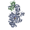
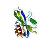
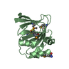
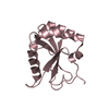
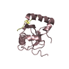
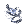
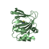
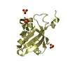
| Title | Structure Of C-Terminal Actin Depolymerizing Factor Homology (Adf-H) Domain Of Human Twinfilin-2 |
|---|
 Components Components | TWINFILIN-2 |
|---|
 Keywords Keywords | TRANSFERASE / CYTOSKELETON / ACTIN-BINDING / ACTIN BINDING / COFILIN-LIKE / PHOSPHOPROTEIN / PHOSPHORYLATION / PROTEIN TYROSINE KINASE-9 / ACTIN DEPOLYMERIZING FACTOR |
|---|
| Function / homology |  Function and homology information Function and homology information
regulation of microvillus length / negative regulation of actin filament polymerization / barbed-end actin filament capping / stereocilium / regulation of lamellipodium assembly / actin filament depolymerization / cell projection organization / Sensory processing of sound by outer hair cells of the cochlea / Sensory processing of sound by inner hair cells of the cochlea / myofibril ...regulation of microvillus length / negative regulation of actin filament polymerization / barbed-end actin filament capping / stereocilium / regulation of lamellipodium assembly / actin filament depolymerization / cell projection organization / Sensory processing of sound by outer hair cells of the cochlea / Sensory processing of sound by inner hair cells of the cochlea / myofibril / actin monomer binding / positive regulation of axon extension / positive regulation of lamellipodium assembly / cellular response to retinoic acid / phosphatidylinositol-4,5-bisphosphate binding / protein kinase C binding / regulation of actin cytoskeleton organization / filopodium / actin filament / positive regulation of neuron projection development / cellular response to growth factor stimulus / actin filament binding / lamellipodium / growth cone / cadherin binding / perinuclear region of cytoplasm / RNA binding / extracellular exosome / ATP binding / cytoplasmSimilarity search - Function Twinfilin / Actin-depolymerising factor homology domain / Cofilin/tropomyosin-type actin-binding protein / ADF-H domain profile. / Actin depolymerisation factor/cofilin -like domains / Severin / Severin / ADF-H/Gelsolin-like domain superfamily / 3-Layer(aba) Sandwich / Alpha BetaSimilarity search - Domain/homology |
|---|
| Biological species |  HOMO SAPIENS (human) HOMO SAPIENS (human) |
|---|
| Method |  X-RAY DIFFRACTION / SYNCHROTRON / MOLECULAR REPLACEMENT / Resolution: 1.8 Å X-RAY DIFFRACTION / SYNCHROTRON / MOLECULAR REPLACEMENT / Resolution: 1.8 Å |
|---|
 Authors Authors | Elkins, J.M. / Pike, A.C.W. / Salah, E. / Savitsky, P. / Murray, J.W. / Roos, A. / von Delft, F. / Edwards, A. / Arrowsmith, C.H. / Wikstrom, M. ...Elkins, J.M. / Pike, A.C.W. / Salah, E. / Savitsky, P. / Murray, J.W. / Roos, A. / von Delft, F. / Edwards, A. / Arrowsmith, C.H. / Wikstrom, M. / Bountra, C. / Knapp, S. |
|---|
 Citation Citation | Journal: To be Published
Title: Structure of C-Terminal Actin Depolymerizing Factor Homology (Adf-H) Domain of Human Twinfilin- 2
Authors: Elkins, J.M. / Pike, A.C.W. / Salah, E. / Savitsky, P. / Murray, J.W. / Roos, A. / von Delft, F. / Edwards, A. / Arrowsmith, C.H. / Wikstrom, M. / Bountra, C. / Knapp, S. |
|---|
| History | | Deposition | Aug 18, 2008 | Deposition site: PDBE / Processing site: PDBE |
|---|
| Revision 1.0 | Sep 30, 2008 | Provider: repository / Type: Initial release |
|---|
| Revision 1.1 | Jul 13, 2011 | Group: Advisory / Version format compliance |
|---|
| Revision 1.2 | Dec 20, 2017 | Group: Database references / Structure summary / Category: audit_author / citation_author / Item: _audit_author.name / _citation_author.name |
|---|
| Revision 1.3 | Dec 13, 2023 | Group: Data collection / Database references ...Data collection / Database references / Other / Refinement description
Category: chem_comp_atom / chem_comp_bond ...chem_comp_atom / chem_comp_bond / database_2 / pdbx_database_status / pdbx_initial_refinement_model
Item: _database_2.pdbx_DOI / _database_2.pdbx_database_accession / _pdbx_database_status.status_code_sf |
|---|
|
|---|
 Movie
Movie Controller

 Yorodumi
 Open data
 Basic information
 Structure visualization
 Downloads & links
 Other downloads


 PDBj
PDBj

 Assembly
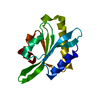

 Mass: 18.015 Da / Num. of mol.: 71 / Source method: isolated from a natural source / Formula: H2O
 Sample preparation
 / Beamline: X10SA / Wavelength: 0.99186
  Processing