 Movie
Movie Controller

[English] 日本語
 Yorodumi
- PDB-1m4j: CRYSTAL STRUCTURE OF THE N-TERMINAL ADF-H DOMAIN OF MOUSE TWINFIL... -
+ Open data

- Basic information
| Entry | Database: PDB / ID: 1m4j | ||||||
|---|---|---|---|---|---|---|---|
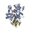
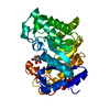
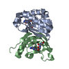
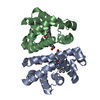
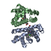
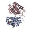
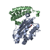
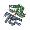
| Title | CRYSTAL STRUCTURE OF THE N-TERMINAL ADF-H DOMAIN OF MOUSE TWINFILIN ISOFORM-1 | ||||||
 Components Components | A6 gene product | ||||||
 Keywords Keywords | STRUCTURAL PROTEIN / MIXED BETA-SHEET / PAIR OF ALPHA-HELICES | ||||||
| Function / homology |  Function and homology information Function and homology informationRHOBTB2 GTPase cycle / negative regulation of actin filament polymerization / barbed-end actin filament capping / actin filament depolymerization / regulation of lamellipodium assembly / positive regulation of cardiac muscle hypertrophy / myofibril / actin monomer binding / phosphatidylinositol-4,5-bisphosphate binding / filopodium ...RHOBTB2 GTPase cycle / negative regulation of actin filament polymerization / barbed-end actin filament capping / actin filament depolymerization / regulation of lamellipodium assembly / positive regulation of cardiac muscle hypertrophy / myofibril / actin monomer binding / phosphatidylinositol-4,5-bisphosphate binding / filopodium / actin filament / positive regulation of neuron projection development / cell-cell junction / actin filament binding / actin cytoskeleton / actin binding / protein tyrosine kinase activity / protein-containing complex binding / perinuclear region of cytoplasm / ATP binding / cytoplasm / cytosol Similarity search - Function | ||||||
| Biological species |  | ||||||
| Method |  X-RAY DIFFRACTION / SYNCHROTRON / MOLECULAR REPLACEMENT / Resolution: 1.6 Å X-RAY DIFFRACTION / SYNCHROTRON / MOLECULAR REPLACEMENT / Resolution: 1.6 Å | ||||||
 Authors Authors | Paavilainen, V.O. / Merckel, M.C. / Falck, S. / Ojala, P.J. / Pohl, E. / Wilmanns, M. / Lappalainen, P. | ||||||
 Citation Citation | Journal: J.Biol.Chem. / Year: 2002 Title: Structural Conservation Between the Actin Monomer-binding Sites of Twinfilin and Actin-depolymerizing Factor (ADF)/Cofilin Authors: Paavilainen, V.O. / Merckel, M.C. / Falck, S. / Ojala, P.J. / Pohl, E. / Wilmanns, M. / Lappalainen, P. #1: Journal: To be PublishedTitle: Twinfilin Forms a Stable Complex with ADP-Actin Monomers Through its Carboxyl-Terminal ADF-H Domain Authors: Ojala, P.J. / Paavilainen, V.O. / Vartiainen, M.K. / Tuma, R. / Weeds, A.G. / Lappalainen, P. #2: Journal: Mol.Cell.Biol. / Year: 2000Title: Mouse A6/twinfilin is an actin monomer-binding protein that localizes to the regions of rapid actin dynamics Authors: Vartiainen, M. / Ojala, P.J. / Auvinen, P. / Peranen, J. / Lappalainen, P. | ||||||
| History |
|
- Structure visualization
| Structure viewer | Molecule: MolmilJmol/JSmol |
|---|
- Downloads & links
-Download
| PDBx/mmCIF format | 1m4j.cif.gz | 65.3 KB | Display | PDBx/mmCIF format |
|---|---|---|---|---|
| PDB format | pdb1m4j.ent.gz | 49.5 KB | Display | PDB format |
| PDBx/mmJSON format | 1m4j.json.gz | Tree view | PDBx/mmJSON format | |
| Others |  Other downloads Other downloads |
-Validation report
| Arichive directory | https://data.pdbj.org/pub/pdb/validation_reports/m4/1m4jftp://data.pdbj.org/pub/pdb/validation_reports/m4/1m4j | HTTPS FTP |
|---|
-Related structure data
| Related structure data | |
|---|---|
| Similar structure data |


-Links
 PDBj
PDBj
- Assembly
| Deposited unit | 
| ||||||||
|---|---|---|---|---|---|---|---|---|---|
| 1 |
| ||||||||
| 2 | 
| ||||||||
| Unit cell |
|
-Components
| #1: Protein | Mass: 16294.358 Da / Num. of mol.: 2 / Fragment: N-terminal ADF-H Domain Source method: isolated from a genetically manipulated source Source: (gene. exp.)  #2: Water | ChemComp-HOH / |  Mass: 18.015 Da / Num. of mol.: 107 / Source method: isolated from a natural source / Formula: H2O Mass: 18.015 Da / Num. of mol.: 107 / Source method: isolated from a natural source / Formula: H2O |
|---|
-Experimental details
-Experiment
| Experiment | Method: X-RAY DIFFRACTION / Number of used crystals: 1 |
|---|
- Sample preparation
| Crystal | Density Matthews: 2.09 Å3/Da / Density % sol: 41.14 % | |||||||||||||||||||||||||||||||||||||||||||||||||
|---|---|---|---|---|---|---|---|---|---|---|---|---|---|---|---|---|---|---|---|---|---|---|---|---|---|---|---|---|---|---|---|---|---|---|---|---|---|---|---|---|---|---|---|---|---|---|---|---|---|---|
| Crystal grow | Temperature: 277 K / Method: vapor diffusion, hanging drop / pH: 7.5 Details: 1.6M Na-Citrate, 3% PEG 400, 0.1M Na-Hepes, pH 7.5, VAPOR DIFFUSION, HANGING DROP, temperature 277K | |||||||||||||||||||||||||||||||||||||||||||||||||
| Crystal grow | *PLUS Temperature: 4 ℃ | |||||||||||||||||||||||||||||||||||||||||||||||||
| Components of the solutions | *PLUS
|
-Data collection
| Diffraction | Mean temperature: 100 K |
|---|---|
| Diffraction source | Source: SYNCHROTRON / Site: EMBL/DESY, HAMBURG  / Beamline: BW7B / Wavelength: 0.8459 Å / Beamline: BW7B / Wavelength: 0.8459 Å |
| Detector | Type: MARRESEARCH / Detector: IMAGE PLATE / Date: Nov 23, 2001 / Details: Premirrors, bent mirror |
| Radiation | Monochromator: Triangular monochromator / Protocol: SINGLE WAVELENGTH / Monochromatic (M) / Laue (L): M / Scattering type: x-ray |
| Radiation wavelength | Wavelength: 0.8459 Å / Relative weight: 1 |
| Reflection | Resolution: 1.6→20 Å / Num. all: 36771 / Num. obs: 36771 / Observed criterion σ(F): 0 / Observed criterion σ(I): 0 / Redundancy: 3.8 % / Biso Wilson estimate: 21.1 Å2 / Rmerge(I) obs: 0.044 / Rsym value: 0.044 |
| Reflection shell | Resolution: 1.6→1.7 Å / Mean I/σ(I) obs: 4 / Num. unique all: 3101 / Rsym value: 0.255 / % possible all: 87.5 |
| Reflection | *PLUS Highest resolution: 1.6 Å / Lowest resolution: 20 Å / Num. obs: 34257 / % possible obs: 93 % / Num. measured all: 457961 |
| Reflection shell | *PLUS Lowest resolution: 1.66 Å / % possible obs: 92.2 % / Rmerge(I) obs: 0.255 |
- Processing
| Software |
| |||||||||||||||||||||||||
|---|---|---|---|---|---|---|---|---|---|---|---|---|---|---|---|---|---|---|---|---|---|---|---|---|---|---|
| Refinement | Method to determine structure: MOLECULAR REPLACEMENT / Resolution: 1.6→20 Å / Rfactor Rfree error: 0.004 / Isotropic thermal model: restrained / Cross valid method: THROUGHOUT / σ(F): 0 / σ(I): 0 / Stereochemistry target values: Engh & Huber / Details: A MODEL BUILT INTO THE SAD MAPS
| |||||||||||||||||||||||||
| Displacement parameters | Biso mean: 21.5 Å2 | |||||||||||||||||||||||||
| Refine analyze |
| |||||||||||||||||||||||||
| Refinement step | Cycle: LAST / Resolution: 1.6→20 Å
| |||||||||||||||||||||||||
| Refine LS restraints |
| |||||||||||||||||||||||||
| LS refinement shell | Resolution: 1.6→1.7 Å / Rfactor Rfree error: 0.012 / Total num. of bins used: 6
| |||||||||||||||||||||||||
| Refinement | *PLUS Lowest resolution: 20 Å / % reflection Rfree: 9 % / Rfactor Rfree: 0.251 / Rfactor Rwork: 0.232 | |||||||||||||||||||||||||
| Solvent computation | *PLUS | |||||||||||||||||||||||||
| Displacement parameters | *PLUS | |||||||||||||||||||||||||
| Refine LS restraints | *PLUS
|
