 Movie
Movie Controller

[English] 日本語
 Yorodumi
- PDB-4qm9: Crystal Structure of a Putative Cysteine Dioxygenase From Bacillu... -
+ Open data

- Basic information
| Entry | Database: PDB / ID: 4qm9 | ||||||
|---|---|---|---|---|---|---|---|
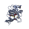
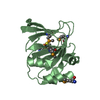
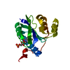
| Title | Crystal Structure of a Putative Cysteine Dioxygenase From Bacillus subtilis with Cys-bound | ||||||
 Components Components | Cysteine dioxygenase | ||||||
 Keywords Keywords | OXIDOREDUCTASE / Cupin fold / catalyzes oxidation of cysteine to cysteine sulfinate | ||||||
| Function / homology |  Function and homology information Function and homology informationcysteine dioxygenase / cysteine dioxygenase activity / oxidoreductase activity, acting on single donors with incorporation of molecular oxygen, incorporation of two atoms of oxygen / ferrous iron binding Similarity search - Function | ||||||
| Biological species |  | ||||||
| Method |  X-RAY DIFFRACTION / SYNCHROTRON / MOLECULAR REPLACEMENT / Resolution: 2.3 Å X-RAY DIFFRACTION / SYNCHROTRON / MOLECULAR REPLACEMENT / Resolution: 2.3 Å | ||||||
 Authors Authors | Hartman, S.H. / Driggers, C.M. / Karplus, P.A. | ||||||
 Citation Citation | Journal: Protein Sci. / Year: 2015 Title: Structures of Arg- and Gln-type bacterial cysteine dioxygenase homologs. Authors: Driggers, C.M. / Hartman, S.J. / Karplus, P.A. | ||||||
| History |
|
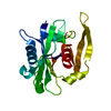
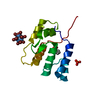
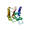
- Structure visualization
| Structure viewer | Molecule: MolmilJmol/JSmol |
|---|
- Downloads & links
-Download
| PDBx/mmCIF format | 4qm9.cif.gz | 137.1 KB | Display | PDBx/mmCIF format |
|---|---|---|---|---|
| PDB format | pdb4qm9.ent.gz | 106.9 KB | Display | PDB format |
| PDBx/mmJSON format | 4qm9.json.gz | Tree view | PDBx/mmJSON format | |
| Others |  Other downloads Other downloads |
-Validation report
| Arichive directory | https://data.pdbj.org/pub/pdb/validation_reports/qm/4qm9ftp://data.pdbj.org/pub/pdb/validation_reports/qm/4qm9 | HTTPS FTP |
|---|
-Related structure data
| Related structure data |  4qm8C  4qmaC 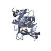 3eqeS S: Starting model for refinement C: citing same article ( |
|---|---|
| Similar structure data |


-Links
 PDBj
PDBj

- Assembly
| Deposited unit | 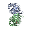
| ||||||||
|---|---|---|---|---|---|---|---|---|---|
| 1 | 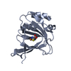
| ||||||||
| 2 | 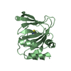
| ||||||||
| Unit cell |
|
-Components
| #1: Protein | Mass: 19614.113 Da / Num. of mol.: 2 Source method: isolated from a genetically manipulated source Source: (gene. exp.) Strain: 168 / Gene: BSU31140, cdoA, Cysteine Dioxygenase, yubC / Plasmid: pET32a / Production host: #2: Chemical |   Mass: 55.845 Da / Num. of mol.: 2 / Source method: obtained synthetically / Formula: Fe Mass: 55.845 Da / Num. of mol.: 2 / Source method: obtained synthetically / Formula: Fe#3: Chemical |   Type: L-peptide linking / Mass: 121.158 Da / Num. of mol.: 2 / Source method: obtained synthetically / Formula: C3H7NO2S Type: L-peptide linking / Mass: 121.158 Da / Num. of mol.: 2 / Source method: obtained synthetically / Formula: C3H7NO2S#4: Water | ChemComp-HOH / |  Mass: 18.015 Da / Num. of mol.: 67 / Source method: isolated from a natural source / Formula: H2O Mass: 18.015 Da / Num. of mol.: 67 / Source method: isolated from a natural source / Formula: H2O |
|---|
-Experimental details
-Experiment
| Experiment | Method: X-RAY DIFFRACTION / Number of used crystals: 1 |
|---|
- Sample preparation
| Crystal | Density Matthews: 2.73 Å3/Da / Density % sol: 54.91 % |
|---|---|
| Crystal grow | Temperature: 298 K / Method: vapor diffusion, hanging drop / pH: 6 Details: Purified enzyme was concentrated to 10 mg/mL and 4 uL was mixed with 4 uL of a reservoir containing 18% (w/v) PEG 4000, 0.1 M potassium acetate, 0.05 M MES, pH 6.0, VAPOR DIFFUSION, HANGING ...Details: Purified enzyme was concentrated to 10 mg/mL and 4 uL was mixed with 4 uL of a reservoir containing 18% (w/v) PEG 4000, 0.1 M potassium acetate, 0.05 M MES, pH 6.0, VAPOR DIFFUSION, HANGING DROP, temperature 298K |
-Data collection
| Diffraction | Mean temperature: 100 K |
|---|---|
| Diffraction source | Source: SYNCHROTRON / Site: ALS  / Beamline: 5.0.1 / Wavelength: 0.976 Å / Beamline: 5.0.1 / Wavelength: 0.976 Å |
| Detector | Type: ADSC QUANTUM 315r / Detector: CCD / Date: Oct 12, 2013 |
| Radiation | Monochromator: Si(220) Asymmetric cut single crystal / Protocol: SINGLE WAVELENGTH / Monochromatic (M) / Laue (L): M / Scattering type: x-ray |
| Radiation wavelength | Wavelength: 0.976 Å / Relative weight: 1 |
| Reflection | Resolution: 2.3→50 Å / Num. all: 20271 / Num. obs: 20271 / % possible obs: 100 % / Observed criterion σ(F): 0 / Observed criterion σ(I): 0 / Redundancy: 23.4 % / Rmerge(I) obs: 0.186 / Rsym value: 0.038 / Net I/σ(I): 13.4 |
| Reflection shell | Resolution: 2.3→2.38 Å / Redundancy: 13.7 % / Rmerge(I) obs: 2.892 / Mean I/σ(I) obs: 1 / Num. unique all: 1939 / Rsym value: 0.767 / % possible all: 100 |
- Processing
| Software |
| ||||||||||||||||||||||||||||||||||||||||||||||||||||||||||||||||||||||||
|---|---|---|---|---|---|---|---|---|---|---|---|---|---|---|---|---|---|---|---|---|---|---|---|---|---|---|---|---|---|---|---|---|---|---|---|---|---|---|---|---|---|---|---|---|---|---|---|---|---|---|---|---|---|---|---|---|---|---|---|---|---|---|---|---|---|---|---|---|---|---|---|---|---|
| Refinement | Method to determine structure: MOLECULAR REPLACEMENT Starting model: 3eqe Resolution: 2.3→46.66 Å / SU ML: 0.41 / σ(F): 1.33 / Phase error: 29.51 / Stereochemistry target values: ML
| ||||||||||||||||||||||||||||||||||||||||||||||||||||||||||||||||||||||||
| Solvent computation | Shrinkage radii: 0.9 Å / VDW probe radii: 1.11 Å / Solvent model: FLAT BULK SOLVENT MODEL | ||||||||||||||||||||||||||||||||||||||||||||||||||||||||||||||||||||||||
| Refinement step | Cycle: LAST / Resolution: 2.3→46.66 Å
| ||||||||||||||||||||||||||||||||||||||||||||||||||||||||||||||||||||||||
| Refine LS restraints |
| ||||||||||||||||||||||||||||||||||||||||||||||||||||||||||||||||||||||||
| LS refinement shell |
| ||||||||||||||||||||||||||||||||||||||||||||||||||||||||||||||||||||||||
| Refinement TLS params. | S33: -0.0256 Å ° / Method: refined / Refine-ID: X-RAY DIFFRACTION
| ||||||||||||||||||||||||||||||||||||||||||||||||||||||||||||||||||||||||
| Refinement TLS group |
|
