 Movie
Movie Controller

[English] 日本語
 Yorodumi
- PDB-2w08: The structure of serum amyloid P component bound to 0-phospho- th... -
+ Open data

- Basic information
| Entry | Database: PDB / ID: 2w08 | ||||||
|---|---|---|---|---|---|---|---|
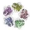
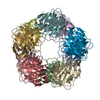
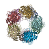
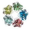
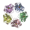
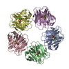
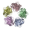
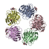
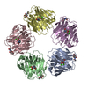
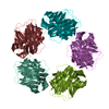
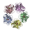
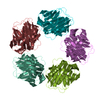
| Title | The structure of serum amyloid P component bound to 0-phospho- threonine | ||||||
 Components Components | SERUM AMYLOID P-COMPONENT | ||||||
 Keywords Keywords | GLYCOPROTEIN / POLYMORPHISM / METAL-BINDING / TAU / LECTIN / CALCIUM / AMYLOID / SECRETED / ALZHEIMERS | ||||||
| Function / homology |  Function and homology information Function and homology informationnegative regulation by host of viral glycoprotein metabolic process / negative regulation of glycoprotein metabolic process / complement component C1q complex binding / negative regulation of viral process / negative regulation of wound healing / negative regulation of monocyte differentiation / host-mediated suppression of symbiont invasion / virion binding / negative regulation of acute inflammatory response / chaperone-mediated protein complex assembly ...negative regulation by host of viral glycoprotein metabolic process / negative regulation of glycoprotein metabolic process / complement component C1q complex binding / negative regulation of viral process / negative regulation of wound healing / negative regulation of monocyte differentiation / host-mediated suppression of symbiont invasion / virion binding / negative regulation of acute inflammatory response / chaperone-mediated protein complex assembly / acute-phase response / : / carbohydrate binding / protein folding / blood microparticle / Amyloid fiber formation / innate immune response / calcium ion binding / : / extracellular exosome / extracellular region / identical protein binding / nucleus Similarity search - Function | ||||||
| Biological species |  HOMO SAPIENS (human) HOMO SAPIENS (human) | ||||||
| Method |  X-RAY DIFFRACTION / SYNCHROTRON / MOLECULAR REPLACEMENT / Resolution: 1.7 Å X-RAY DIFFRACTION / SYNCHROTRON / MOLECULAR REPLACEMENT / Resolution: 1.7 Å | ||||||
 Authors Authors | Kolstoe, S.E. / Pepys, M.B. / Wood, S.P. | ||||||
 Citation Citation | Journal: Proc.Natl.Acad.Sci.USA / Year: 2009 Title: Molecular Dissection of Alzheimer'S Disease Neuropathology by Depletion of Serum Amyloid P Component. Authors: Kolstoe, S.E. / Ridha, B.H. / Bellotti, V. / Wang, N. / Robinson, C.V. / Crutch, S.J. / Keir, G. / Kukkastenvehmas, R. / Gallimore, J.R. / Hutchinson, W.L. / Hawkins, P.N. / Wood, S.P. / ...Authors: Kolstoe, S.E. / Ridha, B.H. / Bellotti, V. / Wang, N. / Robinson, C.V. / Crutch, S.J. / Keir, G. / Kukkastenvehmas, R. / Gallimore, J.R. / Hutchinson, W.L. / Hawkins, P.N. / Wood, S.P. / Rossor, M.N. / Pepys, M.B. | ||||||
| History |
| ||||||
| Remark 700 | SHEET THE SHEET STRUCTURE OF THIS MOLECULE IS BIFURCATED. IN ORDER TO REPRESENT THIS FEATURE IN ... SHEET THE SHEET STRUCTURE OF THIS MOLECULE IS BIFURCATED. IN ORDER TO REPRESENT THIS FEATURE IN THE SHEET RECORDS BELOW, TWO SHEETS ARE DEFINED. |
- Structure visualization
| Structure viewer | Molecule: MolmilJmol/JSmol |
|---|
- Downloads & links
-Download
| PDBx/mmCIF format | 2w08.cif.gz | 250.9 KB | Display | PDBx/mmCIF format |
|---|---|---|---|---|
| PDB format | pdb2w08.ent.gz | 201.9 KB | Display | PDB format |
| PDBx/mmJSON format | 2w08.json.gz | Tree view | PDBx/mmJSON format | |
| Others |  Other downloads Other downloads |
-Validation report
| Arichive directory | https://data.pdbj.org/pub/pdb/validation_reports/w0/2w08ftp://data.pdbj.org/pub/pdb/validation_reports/w0/2w08 | HTTPS FTP |
|---|
-Related structure data
| Related structure data |  1sacS S: Starting model for refinement |
|---|---|
| Similar structure data |


-Links
 PDBj
PDBj
- Assembly
| Deposited unit | 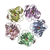
| ||||||||
|---|---|---|---|---|---|---|---|---|---|
| 1 |
| ||||||||
| Unit cell |
|
-Components
| #1: Protein | Mass: 23282.455 Da / Num. of mol.: 5 / Source method: isolated from a natural source / Details: O-PHOSPHOTHREONINE / Source: (natural) HOMO SAPIENS (human) / References: UniProt: P02743#2: Chemical | ChemComp-CA /   Mass: 40.078 Da / Num. of mol.: 10 / Source method: obtained synthetically / Formula: Ca Mass: 40.078 Da / Num. of mol.: 10 / Source method: obtained synthetically / Formula: Ca#3: Sugar | ChemComp-NAG /   Type: D-saccharide, beta linking / Mass: 221.208 Da / Num. of mol.: 5 Type: D-saccharide, beta linking / Mass: 221.208 Da / Num. of mol.: 5Source method: isolated from a genetically manipulated source Formula: C8H15NO6 #4: Chemical | ChemComp-TPO /   Type: L-peptide linking / Mass: 199.099 Da / Num. of mol.: 5 / Source method: obtained synthetically / Formula: C4H10NO6P Type: L-peptide linking / Mass: 199.099 Da / Num. of mol.: 5 / Source method: obtained synthetically / Formula: C4H10NO6P#5: Water | ChemComp-HOH / |  Mass: 18.015 Da / Num. of mol.: 1354 / Source method: isolated from a natural source / Formula: H2O Mass: 18.015 Da / Num. of mol.: 1354 / Source method: isolated from a natural source / Formula: H2OHas protein modification | Y | |
|---|
-Experimental details
-Experiment
| Experiment | Method: X-RAY DIFFRACTION / Number of used crystals: 1 |
|---|
- Sample preparation
| Crystal | Density Matthews: 2.4 Å3/Da / Density % sol: 48.39 % / Description: NONE |
|---|---|
| Crystal grow | pH: 8 Details: 0.06M TRIS-HCL PH8, 16% PEG 550MME, 0.01M CACL2, 0.08M NACL, 0.1% NAN3, 14.2MG/ML PROTEIN, 50MM LIGAND |
-Data collection
| Diffraction | Mean temperature: 100 K |
|---|---|
| Diffraction source | Source: SYNCHROTRON / Site: ESRF  / Beamline: ID14-2 / Wavelength: 0.98 / Beamline: ID14-2 / Wavelength: 0.98 |
| Detector | Type: MARRESEARCH / Detector: CCD / Date: Jul 15, 2004 |
| Radiation | Protocol: SINGLE WAVELENGTH / Monochromatic (M) / Laue (L): M / Scattering type: x-ray |
| Radiation wavelength | Wavelength: 0.98 Å / Relative weight: 1 |
| Reflection | Resolution: 1.7→47.67 Å / Num. obs: 140019 / % possible obs: 97 % / Observed criterion σ(I): 2 / Redundancy: 5.6 % / Rmerge(I) obs: 0.11 / Net I/σ(I): 16.7 |
| Reflection shell | Resolution: 1.7→1.78 Å / Redundancy: 4 % / Rmerge(I) obs: 0.38 / Mean I/σ(I) obs: 9.3 / % possible all: 95.6 |
- Processing
| Software |
| |||||||||||||||||||||||||||||||||||||||||||||||||||||||||||||||||||||||||||||||||||||||||||||||||||||||||
|---|---|---|---|---|---|---|---|---|---|---|---|---|---|---|---|---|---|---|---|---|---|---|---|---|---|---|---|---|---|---|---|---|---|---|---|---|---|---|---|---|---|---|---|---|---|---|---|---|---|---|---|---|---|---|---|---|---|---|---|---|---|---|---|---|---|---|---|---|---|---|---|---|---|---|---|---|---|---|---|---|---|---|---|---|---|---|---|---|---|---|---|---|---|---|---|---|---|---|---|---|---|---|---|---|---|---|
| Refinement | Method to determine structure: MOLECULAR REPLACEMENT Starting model: PDB ENTRY 1SAC Resolution: 1.7→47.48 Å / SU ML: 0.16 / σ(F): 0.04 / Phase error: 15.79 / Stereochemistry target values: ML
| |||||||||||||||||||||||||||||||||||||||||||||||||||||||||||||||||||||||||||||||||||||||||||||||||||||||||
| Solvent computation | Shrinkage radii: 0.9 Å / VDW probe radii: 1.11 Å / Solvent model: FLAT BULK SOLVENT MODEL / Bsol: 47.57 Å2 / ksol: 0.36 e/Å3 | |||||||||||||||||||||||||||||||||||||||||||||||||||||||||||||||||||||||||||||||||||||||||||||||||||||||||
| Refinement step | Cycle: LAST / Resolution: 1.7→47.48 Å
| |||||||||||||||||||||||||||||||||||||||||||||||||||||||||||||||||||||||||||||||||||||||||||||||||||||||||
| Refine LS restraints |
| |||||||||||||||||||||||||||||||||||||||||||||||||||||||||||||||||||||||||||||||||||||||||||||||||||||||||
| LS refinement shell |
|
