#1: Journal: Acta Crystallogr.,Sect.F / Year: 2007 Title: Preliminary Neutron and Ultrahigh-Resolution X-Ray Diffraction Studies of the Aspartic Proteinase Endothiapepsin Cocrystallized with a Gem-Diol Inhibitor. Authors: Tuan, H.-F. / Erskine, P. / Langan, P. / Cooper, J. / Coates, L.
History
Deposition
Apr 17, 2008
Deposition site: PDBE / Processing site: PDBE
Revision 1.0
May 27, 2008
Provider: repository / Type: Initial release
Revision 1.1
Jul 13, 2011
Group: Atomic model / Database references ...Atomic model / Database references / Derived calculations / Non-polymer description / Structure summary / Version format compliance
SHEET THE SHEET STRUCTURE OF THIS MOLECULE IS BIFURCATED. IN ORDER TO REPRESENT THIS FEATURE IN ... SHEET THE SHEET STRUCTURE OF THIS MOLECULE IS BIFURCATED. IN ORDER TO REPRESENT THIS FEATURE IN THE SHEET RECORDS BELOW, TWO SHEETS ARE DEFINED.
In the structure databanks used in Yorodumi, some data are registered as the other names, "COVID-19 virus" and "2019-nCoV". Here are the details of the virus and the list of structure data.
Jan 31, 2019. EMDB accession codes are about to change! (news from PDBe EMDB page)
EMDB accession codes are about to change! (news from PDBe EMDB page)
The allocation of 4 digits for EMDB accession codes will soon come to an end. Whilst these codes will remain in use, new EMDB accession codes will include an additional digit and will expand incrementally as the available range of codes is exhausted. The current 4-digit format prefixed with “EMD-” (i.e. EMD-XXXX) will advance to a 5-digit format (i.e. EMD-XXXXX), and so on. It is currently estimated that the 4-digit codes will be depleted around Spring 2019, at which point the 5-digit format will come into force.
The EM Navigator/Yorodumi systems omit the EMD- prefix.
Related info.:Q: What is EMD? / ID/Accession-code notation in Yorodumi/EM Navigator
Yorodumi is a browser for structure data from EMDB, PDB, SASBDB, etc.
This page is also the successor to EM Navigator detail page, and also detail information page/front-end page for Omokage search.
The word "yorodu" (or yorozu) is an old Japanese word meaning "ten thousand". "mi" (miru) is to see.
Related info.:EMDB / PDB / SASBDB / Comparison of 3 databanks / Yorodumi Search / Aug 31, 2016. New EM Navigator & Yorodumi / Yorodumi Papers / Jmol/JSmol / Function and homology information / Changes in new EM Navigator and Yorodumi
 Movie
Movie Controller

 Yorodumi
 Open data
 Basic information
 Components
 Keywords
 Function and homology information
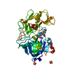
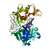
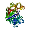
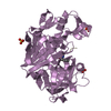
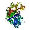
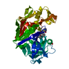
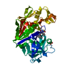
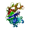
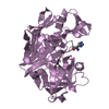
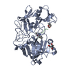
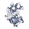
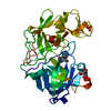
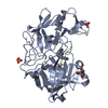
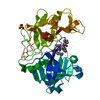
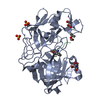
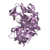
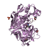
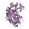
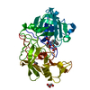
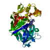
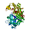
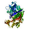
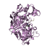
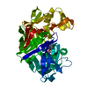
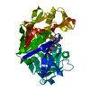
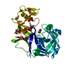
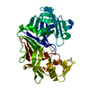
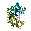
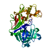
 CRYPHONECTRIA PARASITICA (chestnut blight fungus)
 MOLECULAR REPLACEMENT / Resolution: 2 Å
  Authors
 Citation
 Structure visualization
 Downloads & links
 Other downloads
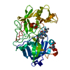


 PDBj
PDBj


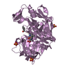
 Assembly
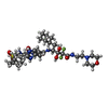
 Type: peptide-like, Peptide-like / Class: Inhibitor / Mass: 784.934 Da / Num. of mol.: 1 / Source method: obtained synthetically / Formula: C36H56F2N7O8S / References: GEM-DIOL INHIBITOR PD-135.040
 Mass: 18.015 Da / Num. of mol.: 220 / Source method: isolated from a natural source / Formula: D2O
 Sample preparation
 / Beamline: MANDI / Wavelength: 1 / Wavelength: 1 Å
 Processing