 Movie
Movie Controller

[English] 日本語
 Yorodumi
- PDB-2voo: Crystal structure of N-terminal domains of Human La protein compl... -
+ Open data

- Basic information
| Entry | Database: PDB / ID: 2voo | ||||||
|---|---|---|---|---|---|---|---|
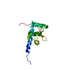
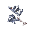
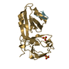
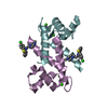
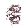
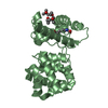
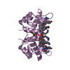
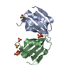
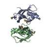
| Title | Crystal structure of N-terminal domains of Human La protein complexed with RNA oligomer UUUUUUUU | ||||||
 Components Components |
| ||||||
 Keywords Keywords | RNA BINDING PROTEIN / RNA-BINDING PROTEIN / RNA RECOGNITION MOTIF / SYSTEMIC LUPUS ERYTHEMATOSUS / PHOSPHOPROTEIN / RNA MATURATION / NUCLEUS / LA MOTIF / RNA-BINDING / POLYMORPHISM | ||||||
| Function / homology |  Function and homology information Function and homology informationnuclear histone mRNA catabolic process / histone mRNA metabolic process / tRNA 3'-end processing / protein localization to cytoplasmic stress granule / IRES-dependent viral translational initiation / RNA Polymerase III Transcription Termination / tRNA modification / tRNA export from nucleus / RNA Polymerase III Abortive And Retractive Initiation / sequence-specific mRNA binding ...nuclear histone mRNA catabolic process / histone mRNA metabolic process / tRNA 3'-end processing / protein localization to cytoplasmic stress granule / IRES-dependent viral translational initiation / RNA Polymerase III Transcription Termination / tRNA modification / tRNA export from nucleus / RNA Polymerase III Abortive And Retractive Initiation / sequence-specific mRNA binding / tRNA 5'-leader removal / tRNA processing / poly(U) RNA binding / positive regulation of translation / cytoplasmic stress granule / chromosome, telomeric region / tRNA binding / ribonucleoprotein complex / mRNA binding / RNA binding / nucleus / cytoplasm / cytosol Similarity search - Function | ||||||
| Biological species |  HOMO SAPIENS (human) HOMO SAPIENS (human) | ||||||
| Method |  X-RAY DIFFRACTION / SYNCHROTRON / MOLECULAR REPLACEMENT / Resolution: 1.8 Å X-RAY DIFFRACTION / SYNCHROTRON / MOLECULAR REPLACEMENT / Resolution: 1.8 Å | ||||||
 Authors Authors | Kotik-Kogan, O. / Valentine, E.R. / Sanfelice, D. / Conte, M.R. / Curry, S. | ||||||
 Citation Citation | Journal: Structure / Year: 2008 Title: Structural Analysis Reveals Conformational Plasticity in the Recognition of RNA 3' Ends by the Human La Protein. Authors: Kotik-Kogan, O. / Valentine, E.R. / Sanfelice, D. / Conte, M.R. / Curry, S. | ||||||
| History |
|
- Structure visualization
| Structure viewer | Molecule: MolmilJmol/JSmol |
|---|
- Downloads & links
-Download
| PDBx/mmCIF format | 2voo.cif.gz | 91.8 KB | Display | PDBx/mmCIF format |
|---|---|---|---|---|
| PDB format | pdb2voo.ent.gz | 68.8 KB | Display | PDB format |
| PDBx/mmJSON format | 2voo.json.gz | Tree view | PDBx/mmJSON format | |
| Others |  Other downloads Other downloads |
-Validation report
| Arichive directory | https://data.pdbj.org/pub/pdb/validation_reports/vo/2vooftp://data.pdbj.org/pub/pdb/validation_reports/vo/2voo | HTTPS FTP |
|---|
-Related structure data
| Related structure data | 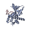 2vodC 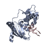 2vonC 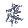 2vopC 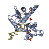 1zh5S S: Starting model for refinement C: citing same article ( |
|---|---|
| Similar structure data |


-Links
 PDBj
PDBj


- Assembly
| Deposited unit | 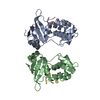
| ||||||||
|---|---|---|---|---|---|---|---|---|---|
| 1 | 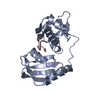
| ||||||||
| 2 | 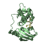
| ||||||||
| Unit cell |
| ||||||||
| Noncrystallographic symmetry (NCS) | NCS oper: (Code: given Matrix: (0.9966, -0.0782, -0.0249), Vector: |
-Components
| #1: Protein | Mass: 22529.809 Da / Num. of mol.: 2 / Fragment: N-TERMINAL DOMAIN, RESIDUES 4-194 Source method: isolated from a genetically manipulated source Source: (gene. exp.) HOMO SAPIENS (human) / Plasmid: PETM11 / Production host:  #2: RNA chain | Mass: 2098.203 Da / Num. of mol.: 2 / Source method: obtained synthetically / Source: (synth.) HOMO SAPIENS (human)#3: Water | ChemComp-HOH / |  Mass: 18.015 Da / Num. of mol.: 201 / Source method: isolated from a natural source / Formula: H2O Mass: 18.015 Da / Num. of mol.: 201 / Source method: isolated from a natural source / Formula: H2OSequence details | FIRST TWO RESIDUES ARE FROM VECTOR (GS) REMAINS, SEQUENCE CORRESPOND | |
|---|
-Experimental details
-Experiment
| Experiment | Method: X-RAY DIFFRACTION / Number of used crystals: 1 |
|---|
- Sample preparation
| Crystal | Density Matthews: 2.33 Å3/Da / Density % sol: 46.81 % / Description: NONE |
|---|
-Data collection
| Diffraction | Mean temperature: 100 K |
|---|---|
| Diffraction source | Source: SYNCHROTRON / Site: ESRF  / Beamline: ID14-1 / Wavelength: 0.62 / Beamline: ID14-1 / Wavelength: 0.62 |
| Detector | Type: ADSC CCD / Detector: CCD / Date: Dec 13, 2006 / Details: SINGLE SILICON (111) MONOCHROMATOR |
| Radiation | Monochromator: SINGLE SILICON (111) MONOCHROMATOR / Protocol: SINGLE WAVELENGTH / Monochromatic (M) / Laue (L): M / Scattering type: x-ray |
| Radiation wavelength | Wavelength: 0.62 Å / Relative weight: 1 |
| Reflection | Resolution: 2.1→41 Å / Num. obs: 3 / % possible obs: 98.7 % / Observed criterion σ(I): 0 / Redundancy: 3.4 % / Biso Wilson estimate: 15.7 Å2 / Rmerge(I) obs: 0.07 / Net I/σ(I): 6.7 |
| Reflection shell | Resolution: 1.8→1.9 Å / Redundancy: 3.4 % / Rmerge(I) obs: 0.04 / Mean I/σ(I) obs: 1.8 / % possible all: 97.7 |
- Processing
| Software |
| ||||||||||||||||||||||||||||||||||||||||||||||||||||||||||||||||||||||||||||||||
|---|---|---|---|---|---|---|---|---|---|---|---|---|---|---|---|---|---|---|---|---|---|---|---|---|---|---|---|---|---|---|---|---|---|---|---|---|---|---|---|---|---|---|---|---|---|---|---|---|---|---|---|---|---|---|---|---|---|---|---|---|---|---|---|---|---|---|---|---|---|---|---|---|---|---|---|---|---|---|---|---|---|
| Refinement | Method to determine structure: MOLECULAR REPLACEMENT Starting model: PDB ENTRY 1ZH5 Resolution: 1.8→37.31 Å / Rfactor Rfree error: 0.006 / Data cutoff high absF: 1715061.49 / Isotropic thermal model: RESTRAINED / Cross valid method: THROUGHOUT / σ(F): 0 / Stereochemistry target values: MLF / Details: BULK SOLVENT MODEL USED
| ||||||||||||||||||||||||||||||||||||||||||||||||||||||||||||||||||||||||||||||||
| Solvent computation | Solvent model: FLAT MODEL / Bsol: 44.0525 Å2 / ksol: 0.4 e/Å3 | ||||||||||||||||||||||||||||||||||||||||||||||||||||||||||||||||||||||||||||||||
| Displacement parameters | Biso mean: 22.2 Å2
| ||||||||||||||||||||||||||||||||||||||||||||||||||||||||||||||||||||||||||||||||
| Refine analyze |
| ||||||||||||||||||||||||||||||||||||||||||||||||||||||||||||||||||||||||||||||||
| Refinement step | Cycle: LAST / Resolution: 1.8→37.31 Å
| ||||||||||||||||||||||||||||||||||||||||||||||||||||||||||||||||||||||||||||||||
| Refine LS restraints |
| ||||||||||||||||||||||||||||||||||||||||||||||||||||||||||||||||||||||||||||||||
| LS refinement shell | Resolution: 1.8→1.91 Å / Rfactor Rfree error: 0.017 / Total num. of bins used: 6
| ||||||||||||||||||||||||||||||||||||||||||||||||||||||||||||||||||||||||||||||||
| Xplor file |
|
