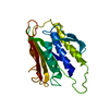
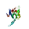
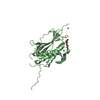
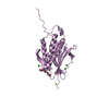
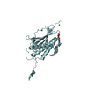
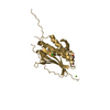
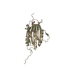
mitotic spindle assembly checkpoint MAD1-MAD2 complex / mitotic checkpoint complex / Inhibition of the proteolytic activity of APC/C required for the onset of anaphase by mitotic spindle checkpoint components / negative regulation of ubiquitin protein ligase activity / Inactivation of APC/C via direct inhibition of the APC/C complex / APC/C:Cdc20 mediated degradation of mitotic proteins / nuclear pore nuclear basket / negative regulation of mitotic cell cycle / positive regulation of mitotic cell cycle spindle assembly checkpoint / mitotic spindle assembly checkpoint signaling ...mitotic spindle assembly checkpoint MAD1-MAD2 complex / mitotic checkpoint complex / Inhibition of the proteolytic activity of APC/C required for the onset of anaphase by mitotic spindle checkpoint components / negative regulation of ubiquitin protein ligase activity / Inactivation of APC/C via direct inhibition of the APC/C complex / APC/C:Cdc20 mediated degradation of mitotic proteins / nuclear pore nuclear basket / negative regulation of mitotic cell cycle / positive regulation of mitotic cell cycle spindle assembly checkpoint / mitotic spindle assembly checkpoint signaling / mitotic sister chromatid segregation / Amplification of signal from unattached kinetochores via a MAD2 inhibitory signal / Mitotic Prometaphase / EML4 and NUDC in mitotic spindle formation / APC-Cdc20 mediated degradation of Nek2A / Resolution of Sister Chromatid Cohesion / Cdc20:Phospho-APC/C mediated degradation of Cyclin A / RHO GTPases Activate Formins / negative regulation of protein catabolic process / kinetochore / spindle pole / mitotic spindle / Separation of Sister Chromatids / cell division / perinuclear region of cytoplasm / protein homodimerization activity / nucleoplasm / identical protein binding / nucleus / cytosol Similarity search - Function
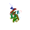
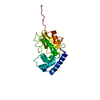
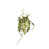
Cell Cycle, Spindle Assembly Checkpoint Protein; Chain A / HORMA domain / Mad2-like / HORMA domain / HORMA domain / HORMA domain profile. / HORMA domain superfamily / 2-Layer Sandwich / Alpha Beta Similarity search - Domain/homology
SHEET THE SHEET STRUCTURE OF THIS MOLECULE IS BIFURCATED. IN ORDER TO REPRESENT THIS FEATURE IN ... SHEET THE SHEET STRUCTURE OF THIS MOLECULE IS BIFURCATED. IN ORDER TO REPRESENT THIS FEATURE IN THE SHEET RECORDS BELOW, TWO SHEETS ARE DEFINED.
Mass: 18.015 Da / Num. of mol.: 1591 / Source method: isolated from a natural source / Formula: H2O
-
Details
Compound details
ENGINEERED RESIDUE IN CHAIN A, LEU 13 TO ALA ENGINEERED RESIDUE IN CHAIN A, CYS 79 TO SER ...ENGINEERED RESIDUE IN CHAIN A, LEU 13 TO ALA ENGINEERED RESIDUE IN CHAIN A, CYS 79 TO SER ENGINEERED RESIDUE IN CHAIN A, CYS 106 TO SER ENGINEERED RESIDUE IN CHAIN B, LEU 13 TO ALA ENGINEERED RESIDUE IN CHAIN B, CYS 79 TO SER ENGINEERED RESIDUE IN CHAIN B, CYS 106 TO SER ENGINEERED RESIDUE IN CHAIN C, LEU 13 TO ALA ENGINEERED RESIDUE IN CHAIN C, CYS 79 TO SER ENGINEERED RESIDUE IN CHAIN C, CYS 106 TO SER ENGINEERED RESIDUE IN CHAIN D, LEU 13 TO ALA ENGINEERED RESIDUE IN CHAIN D, CYS 79 TO SER ENGINEERED RESIDUE IN CHAIN D, CYS 106 TO SER ENGINEERED RESIDUE IN CHAIN E, LEU 13 TO ALA ENGINEERED RESIDUE IN CHAIN E, CYS 79 TO SER ENGINEERED RESIDUE IN CHAIN E, CYS 106 TO SER ENGINEERED RESIDUE IN CHAIN F, LEU 13 TO ALA ENGINEERED RESIDUE IN CHAIN F, CYS 79 TO SER ENGINEERED RESIDUE IN CHAIN F, CYS 106 TO SER ENGINEERED RESIDUE IN CHAIN G, LEU 13 TO ALA ENGINEERED RESIDUE IN CHAIN G, CYS 79 TO SER ENGINEERED RESIDUE IN CHAIN G, CYS 106 TO SER ENGINEERED RESIDUE IN CHAIN H, LEU 13 TO ALA ENGINEERED RESIDUE IN CHAIN H, CYS 79 TO SER ENGINEERED RESIDUE IN CHAIN H, CYS 106 TO SER ENGINEERED RESIDUE IN CHAIN I, LEU 13 TO ALA ENGINEERED RESIDUE IN CHAIN I, CYS 79 TO SER ENGINEERED RESIDUE IN CHAIN I, CYS 106 TO SER ENGINEERED RESIDUE IN CHAIN J, LEU 13 TO ALA ENGINEERED RESIDUE IN CHAIN J, CYS 79 TO SER ENGINEERED RESIDUE IN CHAIN J, CYS 106 TO SER ENGINEERED RESIDUE IN CHAIN K, LEU 13 TO ALA ENGINEERED RESIDUE IN CHAIN K, CYS 79 TO SER ENGINEERED RESIDUE IN CHAIN K, CYS 106 TO SER ENGINEERED RESIDUE IN CHAIN L, LEU 13 TO ALA ENGINEERED RESIDUE IN CHAIN L, CYS 79 TO SER ENGINEERED RESIDUE IN CHAIN L, CYS 106 TO SER
Sequence details
THE FIRST GLYCINE IS PART OF THE CLONING LINKER. L13 HAS BEEN MUTATED TO A
-
Experimental details
-
Experiment
Experiment
Method: X-RAY DIFFRACTION / Number of used crystals: 1
-
Sample preparation
Crystal
Density Matthews: 2.9 Å3/Da / Density % sol: 57.1 % / Description: NONE
Crystal grow
Temperature: 293 K / Method: vapor diffusion, hanging drop / pH: 8 Details: VAPOR DIFFUSION, HANGING DROP, 20 DEGREES C. 1 MICROLITER PROTEIN: 3 MG/ML IN 20 MM TRIS, PH 8.0, 50 MM NACL, 2 MM TCEP PLUS 1 MICROLITER RESERVOIR: 19% PEG2000, 16% GLYCEROL, 100 MM TRIS, PH 8.0, 0.3 M MGCL2
 Movie
Movie Controller


 Open data
 Basic information
 Components
 Keywords
 Function and homology information
 HOMO SAPIENS (human)
 X-RAY DIFFRACTION /
  Authors
 Citation
 Structure visualization
 Downloads & links
 Other downloads


 PDBj
PDBj


 Assembly


 Mass: 24.305 Da / Num. of mol.: 8 / Source method: obtained synthetically / Formula: Mg
 Mass: 35.453 Da / Num. of mol.: 37 / Source method: obtained synthetically / Formula: Cl
 Mass: 354.436 Da / Num. of mol.: 3 / Source method: obtained synthetically / Formula: C16H34O8 / Comment: precipitant*YM
 Mass: 634.751 Da / Num. of mol.: 6 / Source method: obtained synthetically / Formula: C28H58O15
 Mass: 106.120 Da / Num. of mol.: 1 / Source method: obtained synthetically / Formula: C4H10O3
 Sample preparation
 / Beamline: 19-ID / Wavelength: 0.97929
  Processing