 Movie
Movie Controller

[English] 日本語
 Yorodumi
- PDB-1vst: Symmetric Sulfolobus solfataricus uracil phosphoribosyltransferas... -
+ Open data

- Basic information
| Entry | Database: PDB / ID: 1vst | ||||||
|---|---|---|---|---|---|---|---|
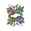
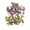
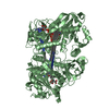
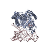
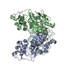
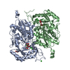
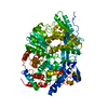
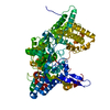
| Title | Symmetric Sulfolobus solfataricus uracil phosphoribosyltransferase with bound PRPP and GTP | ||||||
 Components Components | Uracil phosphoribosyltransferase | ||||||
 Keywords Keywords | TRANSFERASE / uracil / phosphoribosyltransferase / allosteric regulation / Sulfolobus solfataricus / PRPP / GTP / Glycosyltransferase / Magnesium | ||||||
| Function / homology |  Function and homology information Function and homology informationuracil salvage / uracil phosphoribosyltransferase / uracil phosphoribosyltransferase activity / guanine salvage / hypoxanthine metabolic process / hypoxanthine phosphoribosyltransferase activity / UMP salvage / GMP salvage / IMP salvage / GTP binding ...uracil salvage / uracil phosphoribosyltransferase / uracil phosphoribosyltransferase activity / guanine salvage / hypoxanthine metabolic process / hypoxanthine phosphoribosyltransferase activity / UMP salvage / GMP salvage / IMP salvage / GTP binding / magnesium ion binding / cytosol Similarity search - Function | ||||||
| Biological species |   Sulfolobus solfataricus (archaea) Sulfolobus solfataricus (archaea) | ||||||
| Method |  X-RAY DIFFRACTION / SYNCHROTRON / MOLECULAR REPLACEMENT / Resolution: 2.8 Å X-RAY DIFFRACTION / SYNCHROTRON / MOLECULAR REPLACEMENT / Resolution: 2.8 Å | ||||||
 Authors Authors | Kadziola, A. | ||||||
 Citation Citation | Journal: J.Mol.Biol. / Year: 2009 Title: Structural and kinetic studies of the allosteric transition in Sulfolobus solfataricus uracil phosphoribosyltransferase: Permanent activation by engineering of the C-terminus Authors: Christoffersen, S. / Kadziola, A. / Johansson, E. / Rasmussen, M. / Willemoes, M. / Jensen, K.F. #1: Journal: Biochemistry / Year: 2005Title: Allosteric regulation and communication between subunits in uracil phosphoribosyltransferase from Sulfolobus solfataricus. Authors: Arent, S. / Harris, P. / Jensen, K.F. / Larsen, S. | ||||||
| History |
|
- Structure visualization
| Structure viewer | Molecule: MolmilJmol/JSmol |
|---|
- Downloads & links
-Download
| PDBx/mmCIF format | 1vst.cif.gz | 57.1 KB | Display | PDBx/mmCIF format |
|---|---|---|---|---|
| PDB format | pdb1vst.ent.gz | 41.1 KB | Display | PDB format |
| PDBx/mmJSON format | 1vst.json.gz | Tree view | PDBx/mmJSON format | |
| Others |  Other downloads Other downloads |
-Validation report
| Arichive directory | https://data.pdbj.org/pub/pdb/validation_reports/vs/1vstftp://data.pdbj.org/pub/pdb/validation_reports/vs/1vst | HTTPS FTP |
|---|
-Related structure data
| Related structure data |  3g6wC 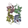 1xtvS S: Starting model for refinement C: citing same article ( |
|---|---|
| Similar structure data |


-Links
 PDBj
PDBj
- Assembly
| Deposited unit | 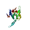
| ||||||||
|---|---|---|---|---|---|---|---|---|---|
| 1 | 
| ||||||||
| Unit cell |
| ||||||||
| Components on special symmetry positions |
|
-Components
| #1: Protein | Mass: 24179.348 Da / Num. of mol.: 1 Source method: isolated from a genetically manipulated source Source: (gene. exp.) Sulfolobus solfataricus (archaea) / Gene: SSO0231, upp / Plasmid: pLFS2 / Production host:  References: UniProt: Q980Q4, uracil phosphoribosyltransferase |
|---|---|
| #2: Chemical | ChemComp-GTP /   Mass: 523.180 Da / Num. of mol.: 1 / Source method: obtained synthetically / Formula: C10H16N5O14P3 / Comment: GTP, energy-carrying molecule*YM Mass: 523.180 Da / Num. of mol.: 1 / Source method: obtained synthetically / Formula: C10H16N5O14P3 / Comment: GTP, energy-carrying molecule*YM |
| #3: Chemical | ChemComp-MG /   Mass: 24.305 Da / Num. of mol.: 1 / Source method: obtained synthetically / Formula: Mg Mass: 24.305 Da / Num. of mol.: 1 / Source method: obtained synthetically / Formula: Mg |
| #4: Sugar | ChemComp-PRP /   Type: D-saccharide / Mass: 390.070 Da / Num. of mol.: 1 / Source method: obtained synthetically / Formula: C5H13O14P3 Type: D-saccharide / Mass: 390.070 Da / Num. of mol.: 1 / Source method: obtained synthetically / Formula: C5H13O14P3 |
| #5: Water | ChemComp-HOH /  Mass: 18.015 Da / Num. of mol.: 10 / Source method: isolated from a natural source / Formula: H2O Mass: 18.015 Da / Num. of mol.: 10 / Source method: isolated from a natural source / Formula: H2O |
-Experimental details
-Experiment
| Experiment | Method: X-RAY DIFFRACTION / Number of used crystals: 1 |
|---|
- Sample preparation
| Crystal | Density Matthews: 2.77 Å3/Da / Density % sol: 55.63 % |
|---|---|
| Crystal grow | Temperature: 298 K / Method: vapor diffusion, hanging drop / pH: 6.5 Details: 10% PEG8000, 0.1M sodium-cacodylate, pH6.5, 200mM magnesium-chloride, 10% glycerol, VAPOR DIFFUSION, HANGING DROP, temperature 298K |
-Data collection
| Diffraction | Mean temperature: 120 K |
|---|---|
| Diffraction source | Source: SYNCHROTRON / Site: MAX II  / Beamline: I711 / Wavelength: 0.954 Å / Beamline: I711 / Wavelength: 0.954 Å |
| Detector | Type: MAR CCD 165 mm / Detector: CCD / Date: Feb 22, 2005 |
| Radiation | Monochromator: GRAPHITE / Protocol: SINGLE WAVELENGTH / Monochromatic (M) / Laue (L): M / Scattering type: x-ray |
| Radiation wavelength | Wavelength: 0.954 Å / Relative weight: 1 |
| Reflection | Resolution: 2.8→25 Å / Num. all: 7102 / Num. obs: 7102 / % possible obs: 99.9 % / Redundancy: 9.7 % / Rmerge(I) obs: 0.152 / Net I/σ(I): 15.9 |
| Reflection shell | Resolution: 2.8→2.87 Å / Rmerge(I) obs: 0.418 / Mean I/σ(I) obs: 4.2 / % possible all: 99.3 |
- Processing
| Software |
| |||||||||||||||||||||||||
|---|---|---|---|---|---|---|---|---|---|---|---|---|---|---|---|---|---|---|---|---|---|---|---|---|---|---|
| Refinement | Method to determine structure: MOLECULAR REPLACEMENT Starting model: PDB ENTRY 1XTV Resolution: 2.8→25 Å / Isotropic thermal model: isotropic / Cross valid method: R-free / Stereochemistry target values: Engh & Huber
| |||||||||||||||||||||||||
| Displacement parameters | Biso mean: 20 Å2
| |||||||||||||||||||||||||
| Refine analyze |
| |||||||||||||||||||||||||
| Refinement step | Cycle: LAST / Resolution: 2.8→25 Å
| |||||||||||||||||||||||||
| Refine LS restraints |
|
