Protein / Protein/peptide , 2 types, 8 molecules ABCDEFGH
#1: Protein
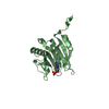
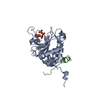
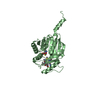
PutativeSAM-dependentmethyltransferase
Mass: 28509.682 Da / Num. of mol.: 4 Source method: isolated from a genetically manipulated source Source: (gene. exp.) Streptomyces lasaliensis (bacteria) / Gene: ecm18 / Plasmid: pKW469 / Production host: Escherichia coli (E. coli) / Strain (production host): BL21(DE3) / References: UniProt: Q0X0A7
#2: Protein/peptide
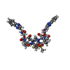
Echinomycin
Type: Cyclic depsipeptide / Class: Antibiotic / Mass: 809.008 Da / Num. of mol.: 4 / Source method: obtained synthetically Details: ECHINOMYCIN IS A BICYCLIC OCTADEPSIPEPTIDE. BICYCLIZATION IS ACHIEVED BY LINKING THE N- AND THE C- TERMINI, AND A THIOACETAL BOND BETWEEN RESIDUES 3 AND 7. THE TWO QUINOXALINE CHROMOPHORES ...Details: ECHINOMYCIN IS A BICYCLIC OCTADEPSIPEPTIDE. BICYCLIZATION IS ACHIEVED BY LINKING THE N- AND THE C- TERMINI, AND A THIOACETAL BOND BETWEEN RESIDUES 3 AND 7. THE TWO QUINOXALINE CHROMOPHORES ARE LINKED TO THE D-SERINE RESIDUES, RESIDUES 1 AND 5. Source: (synth.) Streptomyces echinatus (bacteria) / References: NOR: NOR01126, Echinomycin
Type: Cyclic depsipeptide / Class: Antibiotic / Mass: 174.156 Da / Num. of mol.: 8 / Source method: obtained synthetically / Formula: C9H6N2O2 Details: ECHINOMYCIN IS A BICYCLIC OCTADEPSIPEPTIDE. BICYCLIZATION IS ACHIEVED BY LINKING THE N- AND THE C- TERMINI, AND A THIOACETAL BOND BETWEEN RESIDUES 3 AND 7. THE TWO QUINOXALINE CHROMOPHORES ...Details: ECHINOMYCIN IS A BICYCLIC OCTADEPSIPEPTIDE. BICYCLIZATION IS ACHIEVED BY LINKING THE N- AND THE C- TERMINI, AND A THIOACETAL BOND BETWEEN RESIDUES 3 AND 7. THE TWO QUINOXALINE CHROMOPHORES ARE LINKED TO THE D-SERINE RESIDUES, RESIDUES 1 AND 5. References: Echinomycin
Mass: 18.015 Da / Num. of mol.: 320 / Source method: isolated from a natural source / Formula: H2O
-
Details
Compound details
THE ECHINOMYCIN IS A BICYCLIC OCTADEPSIPEPTIDE, A MEMBER OF THE QUINOXALINE CLASS OF ANTIBIOTICS. ...THE ECHINOMYCIN IS A BICYCLIC OCTADEPSIPEPTIDE, A MEMBER OF THE QUINOXALINE CLASS OF ANTIBIOTICS. HERE, ECHINOMYCIN IS REPRESENTED BY GROUPING TOGETHER THE SEQUENCE (SEQRES) AND TWO LIGANDS (HET) QUI.
-
Experimental details
-
Experiment
Experiment
Method: X-RAY DIFFRACTION / Number of used crystals: 1
-
Sample preparation
Crystal
Density Matthews: 2.07 Å3/Da / Density % sol: 40.55 %
 Movie
Movie Controller

 Yorodumi
 Open data
 Basic information
 Components
 Keywords
 Function and homology information
 Streptomyces lasaliensis (bacteria)
 X-RAY DIFFRACTION /
  Authors
 Citation
 Structure visualization
 Downloads & links
 Other downloads
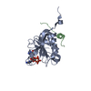


 PDBj
PDBj Assembly


 Type: Cyclic depsipeptide / Class: Antibiotic / Mass: 809.008 Da / Num. of mol.: 4 / Source method: obtained synthetically


 Type: L-peptide linking / Mass: 384.411 Da / Num. of mol.: 4 / Source method: obtained synthetically / Formula: C14H20N6O5S
 Mass: 150.173 Da / Num. of mol.: 2 / Source method: obtained synthetically / Formula: C6H14O4
 Mass: 22.990 Da / Num. of mol.: 3 / Source method: obtained synthetically / Formula: Na
 Type: Cyclic depsipeptide / Class: Antibiotic / Mass: 174.156 Da / Num. of mol.: 8 / Source method: obtained synthetically / Formula: C9H6N2O2
 Mass: 59.044 Da / Num. of mol.: 1 / Source method: obtained synthetically / Formula: C2H3O2
 Mass: 1529.829 Da / Num. of mol.: 2 / Source method: obtained synthetically / Formula: C69H140O35 / Comment: precipitant*YM
 Sample preparation
 / Beamline: 17-ID / Wavelength: 1 Å
 Processing