 Movie
Movie Controller

[English] 日本語
 Yorodumi
- PDB-4pwp: Crystal structure of DsbA from the Gram positive bacterium Coryne... -
+ Open data

- Basic information
| Entry | Database: PDB / ID: 4pwp | ||||||
|---|---|---|---|---|---|---|---|
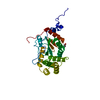
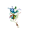
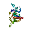
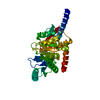
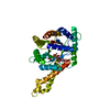
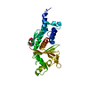
| Title | Crystal structure of DsbA from the Gram positive bacterium Corynebacterium diphtheriae | ||||||
 Components Components | DsbA | ||||||
 Keywords Keywords | STRUCTURAL GENOMICS / thioredoxin domain / disulfide bond isomerase | ||||||
| Function / homology | : / DsbA, N-terminal domain / Thioredoxin / Thioredoxin-like fold / Thioredoxin domain profile. / Thioredoxin domain / Thioredoxin-like superfamily / oxidoreductase activity / Secreted protein Function and homology information Function and homology information | ||||||
| Biological species |  Corynebacterium diphtheriae (bacteria) Corynebacterium diphtheriae (bacteria) | ||||||
| Method |  X-RAY DIFFRACTION / SYNCHROTRON / SAD / Resolution: 1.803 Å X-RAY DIFFRACTION / SYNCHROTRON / SAD / Resolution: 1.803 Å | ||||||
 Authors Authors | Um, S.H. / Kim, J.S. / Jiao, L. / Yoon, B.Y. / Jo, I. / Ha, N.C. | ||||||
 Citation Citation | Journal: To be Published Title: Crystal structure and biochemical characterization of DsbA from the Gram positive bacterium Corynebacterium diphtheriae Authors: Um, S.H. / Kim, J.S. / Jiao, L. / Yoon, B.Y. / Jo, I. / Ha, N.C. | ||||||
| History |
|
- Structure visualization
| Structure viewer | Molecule: MolmilJmol/JSmol |
|---|
- Downloads & links
-Download
| PDBx/mmCIF format | 4pwp.cif.gz | 67.7 KB | Display | PDBx/mmCIF format |
|---|---|---|---|---|
| PDB format | pdb4pwp.ent.gz | 49.8 KB | Display | PDB format |
| PDBx/mmJSON format | 4pwp.json.gz | Tree view | PDBx/mmJSON format | |
| Others |  Other downloads Other downloads |
-Validation report
| Arichive directory | https://data.pdbj.org/pub/pdb/validation_reports/pw/4pwpftp://data.pdbj.org/pub/pdb/validation_reports/pw/4pwp | HTTPS FTP |
|---|
-Related structure data


-Links
 PDBj
PDBj- Assembly
| Deposited unit | 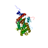
| ||||||||
|---|---|---|---|---|---|---|---|---|---|
| 1 |
| ||||||||
| Unit cell |
|
-Components
| #1: Protein | Mass: 27047.551 Da / Num. of mol.: 1 Source method: isolated from a genetically manipulated source Source: (gene. exp.) Corynebacterium diphtheriae (bacteria) / Production host: |
|---|---|
| #2: Chemical | ChemComp-GOL /   Mass: 92.094 Da / Num. of mol.: 1 / Source method: obtained synthetically / Formula: C3H8O3 Mass: 92.094 Da / Num. of mol.: 1 / Source method: obtained synthetically / Formula: C3H8O3 |
| #3: Water | ChemComp-HOH /  Mass: 18.015 Da / Num. of mol.: 231 / Source method: isolated from a natural source / Formula: H2O Mass: 18.015 Da / Num. of mol.: 231 / Source method: isolated from a natural source / Formula: H2O |
| Has protein modification | Y |
| Sequence details | THE SEQUENCE OF THIS PROTEIN WAS NOT AVAILABLE AT THE UNIPROT KNOWLEDGEBASE DATABASE (UNIPROTKB) AT ...THE SEQUENCE OF THIS PROTEIN WAS NOT AVAILABLE AT THE UNIPROT KNOWLEDGEB |
-Experimental details
-Experiment
| Experiment | Method: X-RAY DIFFRACTION / Number of used crystals: 1 |
|---|
- Sample preparation
| Crystal | Density Matthews: 2.14 Å3/Da / Density % sol: 42.39 % |
|---|---|
| Crystal grow | Temperature: 287 K / Method: vapor diffusion, hanging drop / pH: 8 Details: 30% PEG 2K, pH 8.0, VAPOR DIFFUSION, HANGING DROP, temperature 287K |
-Data collection
| Diffraction | Mean temperature: 100 K | |||||||||||||||||||||
|---|---|---|---|---|---|---|---|---|---|---|---|---|---|---|---|---|---|---|---|---|---|---|
| Diffraction source | Source: SYNCHROTRON / Site: PAL/PLS  / Beamline: 5C (4A) / Wavelength: 1 Å / Beamline: 5C (4A) / Wavelength: 1 Å | |||||||||||||||||||||
| Detector | Type: ADSC QUANTUM 315r / Detector: CCD / Date: Jun 26, 2013 | |||||||||||||||||||||
| Radiation | Monochromator: double mirrors / Protocol: SINGLE WAVELENGTH / Monochromatic (M) / Laue (L): M / Scattering type: x-ray | |||||||||||||||||||||
| Radiation wavelength | Wavelength: 1 Å / Relative weight: 1 | |||||||||||||||||||||
| Reflection | Resolution: 1.48→50 Å / Num. all: 41100 / Num. obs: 40982 / % possible obs: 99.7 % / Observed criterion σ(F): 0 / Observed criterion σ(I): 0 | |||||||||||||||||||||
| Reflection shell |
|
- Processing
| Software |
| ||||||||||||||||||||||||||||||||||||||||||||||||||||||||||||||||||||||||||||||||||||||||||
|---|---|---|---|---|---|---|---|---|---|---|---|---|---|---|---|---|---|---|---|---|---|---|---|---|---|---|---|---|---|---|---|---|---|---|---|---|---|---|---|---|---|---|---|---|---|---|---|---|---|---|---|---|---|---|---|---|---|---|---|---|---|---|---|---|---|---|---|---|---|---|---|---|---|---|---|---|---|---|---|---|---|---|---|---|---|---|---|---|---|---|---|
| Refinement | Method to determine structure: SAD / Resolution: 1.803→30.856 Å / SU ML: 0.16 / σ(F): 0.93 / Phase error: 17.98 / Stereochemistry target values: MLHL
| ||||||||||||||||||||||||||||||||||||||||||||||||||||||||||||||||||||||||||||||||||||||||||
| Solvent computation | Shrinkage radii: 0.9 Å / VDW probe radii: 1.11 Å / Solvent model: FLAT BULK SOLVENT MODEL | ||||||||||||||||||||||||||||||||||||||||||||||||||||||||||||||||||||||||||||||||||||||||||
| Refinement step | Cycle: LAST / Resolution: 1.803→30.856 Å
| ||||||||||||||||||||||||||||||||||||||||||||||||||||||||||||||||||||||||||||||||||||||||||
| Refine LS restraints |
| ||||||||||||||||||||||||||||||||||||||||||||||||||||||||||||||||||||||||||||||||||||||||||
| LS refinement shell | Refine-ID: X-RAY DIFFRACTION / Total num. of bins used: 14
|
