 Movie
Movie Controller

[English] 日本語
 Yorodumi
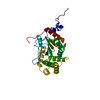
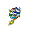
- PDB-5e05: Structure of Sin Nombre virus nucleoprotein in shot-axis crystal form -
+ Open data

- Basic information
| Entry | Database: PDB / ID: 500000 | ||||||
|---|---|---|---|---|---|---|---|
| Title | Structure of Sin Nombre virus nucleoprotein in shot-axis crystal form | ||||||
 Components Components | Nucleocapsid protein | ||||||
 Keywords Keywords | NUCLEAR PROTEIN / hantavirus / Sin Nombre virus / nucleoprotein | ||||||
| Function / homology |  Function and homology information Function and homology informationviral nucleocapsid / endonuclease activity / Hydrolases; Acting on ester bonds / host cell Golgi apparatus / host cell perinuclear region of cytoplasm / ribonucleoprotein complex / hydrolase activity / RNA binding Similarity search - Function | ||||||
| Biological species |  Sin Nombre virus Sin Nombre virus | ||||||
| Method |  X-RAY DIFFRACTION / SYNCHROTRON / MOLECULAR REPLACEMENT / Resolution: 2.3 Å X-RAY DIFFRACTION / SYNCHROTRON / MOLECULAR REPLACEMENT / Resolution: 2.3 Å | ||||||
 Authors Authors | Guo, Y. / Wang, W.M. / Lou, Z.Y. | ||||||
 Citation Citation | Journal: J.Virol. / Year: 2015 Title: Crystal Structure of the Core Region of Hantavirus Nucleocapsid Protein Reveals the Mechanism for Ribonucleoprotein Complex Formation Authors: Guo, Y. / Wang, W. / Sun, Y. / Ma, C. / Wang, X. / Wang, X. / Liu, P. / Shen, S. / Li, B. / Lin, J. / Deng, F. / Wang, H. / Lou, Z. | ||||||
| History |
|
- Structure visualization
| Structure viewer | Molecule: MolmilJmol/JSmol |
|---|
- Downloads & links
-Download
| PDBx/mmCIF format | 5e05.cif.gz | 121.3 KB | Display | PDBx/mmCIF format |
|---|---|---|---|---|
| PDB format | pdb5e05.ent.gz | 93.8 KB | Display | PDB format |
| PDBx/mmJSON format | 5e05.json.gz | Tree view | PDBx/mmJSON format | |
| Others |  Other downloads Other downloads |
-Validation report
| Arichive directory | https://data.pdbj.org/pub/pdb/validation_reports/e0/5e05ftp://data.pdbj.org/pub/pdb/validation_reports/e0/5e05 | HTTPS FTP |
|---|
-Related structure data
| Related structure data |  5e04C 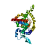 5e06SC  5e07 C: citing same article ( S: Starting model for refinement |
|---|---|
| Similar structure data |


-Links
 PDBj
PDBj- Assembly
| Deposited unit | 
| ||||||||
|---|---|---|---|---|---|---|---|---|---|
| 1 |
| ||||||||
| Unit cell |
|
-Components
| #1: Protein | Mass: 32158.184 Da / Num. of mol.: 1 / Fragment: UNP residues 112-395 Source method: isolated from a genetically manipulated source Source: (gene. exp.) Sin Nombre virus / Production host:  |
|---|---|
| #2: Chemical | ChemComp-PO4 /   Mass: 94.971 Da / Num. of mol.: 1 / Source method: obtained synthetically / Formula: PO4 Mass: 94.971 Da / Num. of mol.: 1 / Source method: obtained synthetically / Formula: PO4 |
| #3: Water | ChemComp-HOH /  Mass: 18.015 Da / Num. of mol.: 45 / Source method: isolated from a natural source / Formula: H2O Mass: 18.015 Da / Num. of mol.: 45 / Source method: isolated from a natural source / Formula: H2O |
-Experimental details
-Experiment
| Experiment | Method: X-RAY DIFFRACTION |
|---|
- Sample preparation
| Crystal | Density Matthews: 2.24 Å3/Da / Density % sol: 44.98 % |
|---|---|
| Crystal grow | Temperature: 289 K / Method: vapor diffusion, hanging drop Details: 200 mM HEPES (pH 7.5), 200 mM lithium sulfate monohydrate, 23% (w/v) PEG3350 |
-Data collection
| Diffraction | Mean temperature: 100 K |
|---|---|
| Diffraction source | Source: SYNCHROTRON / Site: SSRF  / Beamline: BL17U / Wavelength: 0.9798 Å / Beamline: BL17U / Wavelength: 0.9798 Å |
| Detector | Type: ADSC QUANTUM 315 / Detector: CCD / Date: Jun 17, 2014 |
| Radiation | Protocol: SINGLE WAVELENGTH / Monochromatic (M) / Laue (L): M / Scattering type: x-ray |
| Radiation wavelength | Wavelength: 0.9798 Å / Relative weight: 1 |
| Reflection | Resolution: 2.3→50 Å / Num. obs: 13234 / % possible obs: 100 % / Redundancy: 7.8 % / Net I/σ(I): 31 |
- Processing
| Software |
| ||||||||||||||||||||||||||||||||||||||||||||||||||||||||||||||||||||||||||||||||||||||||||||||||||||
|---|---|---|---|---|---|---|---|---|---|---|---|---|---|---|---|---|---|---|---|---|---|---|---|---|---|---|---|---|---|---|---|---|---|---|---|---|---|---|---|---|---|---|---|---|---|---|---|---|---|---|---|---|---|---|---|---|---|---|---|---|---|---|---|---|---|---|---|---|---|---|---|---|---|---|---|---|---|---|---|---|---|---|---|---|---|---|---|---|---|---|---|---|---|---|---|---|---|---|---|---|---|
| Refinement | Method to determine structure: MOLECULAR REPLACEMENT / Starting model: 5000000 / Resolution: 2.3→41.438 Å / SU ML: 0.21 / Cross valid method: FREE R-VALUE / σ(F): 1.34 / Phase error: 23.09 / Stereochemistry target values: ML
| ||||||||||||||||||||||||||||||||||||||||||||||||||||||||||||||||||||||||||||||||||||||||||||||||||||
| Solvent computation | Shrinkage radii: 0.9 Å / VDW probe radii: 1.11 Å / Solvent model: FLAT BULK SOLVENT MODEL | ||||||||||||||||||||||||||||||||||||||||||||||||||||||||||||||||||||||||||||||||||||||||||||||||||||
| Refinement step | Cycle: LAST / Resolution: 2.3→41.438 Å
| ||||||||||||||||||||||||||||||||||||||||||||||||||||||||||||||||||||||||||||||||||||||||||||||||||||
| Refine LS restraints |
| ||||||||||||||||||||||||||||||||||||||||||||||||||||||||||||||||||||||||||||||||||||||||||||||||||||
| LS refinement shell |
| ||||||||||||||||||||||||||||||||||||||||||||||||||||||||||||||||||||||||||||||||||||||||||||||||||||
| Refinement TLS params. | Method: refined / Refine-ID: X-RAY DIFFRACTION
| ||||||||||||||||||||||||||||||||||||||||||||||||||||||||||||||||||||||||||||||||||||||||||||||||||||
| Refinement TLS group |
|
