 Movie
Movie Controller


+ Open data

- Basic information
| Entry | Database: PDB / ID: 6hut | ||||||
|---|---|---|---|---|---|---|---|
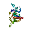
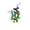
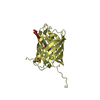
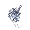
| Title | GFP8 - a stabilized variant of cycle-3 GFP | ||||||
 Components Components | Green fluorescent protein | ||||||
 Keywords Keywords | FLUORESCENT PROTEIN / green fluorescent protein / cycle-3 | ||||||
| Function / homology | Green fluorescent protein, GFP / Green fluorescent protein-related / Green fluorescent protein / Green fluorescent protein / bioluminescence / generation of precursor metabolites and energy / Green fluorescent protein / Green fluorescent protein Function and homology information Function and homology information | ||||||
| Biological species |   Aequorea victoria (jellyfish) Aequorea victoria (jellyfish) | ||||||
| Method |  X-RAY DIFFRACTION / SYNCHROTRON / MOLECULAR REPLACEMENT / Resolution: 1.29 Å X-RAY DIFFRACTION / SYNCHROTRON / MOLECULAR REPLACEMENT / Resolution: 1.29 Å | ||||||
 Authors Authors | Petek, M. / Rangel Pereira, M. | ||||||
| Funding support |  United Kingdom, 1items United Kingdom, 1items
| ||||||
 Citation Citation | Journal: To Be Published Title: GFP8 - a stabilized variant of cycle-3 GFP Authors: Petek, M. / Rangel Pereira, M. | ||||||
| History |
|
- Structure visualization
| Structure viewer | Molecule: MolmilJmol/JSmol |
|---|
- Downloads & links
-Download
| PDBx/mmCIF format | 6hut.cif.gz | 103.4 KB | Display | PDBx/mmCIF format |
|---|---|---|---|---|
| PDB format | pdb6hut.ent.gz | 79 KB | Display | PDB format |
| PDBx/mmJSON format | 6hut.json.gz | Tree view | PDBx/mmJSON format | |
| Others |  Other downloads Other downloads |
-Validation report
| Arichive directory | https://data.pdbj.org/pub/pdb/validation_reports/hu/6hutftp://data.pdbj.org/pub/pdb/validation_reports/hu/6hut | HTTPS FTP |
|---|
-Related structure data
| Related structure data |  4lqtS S: Starting model for refinement |
|---|---|
| Similar structure data |


-Links
 PDBj
PDBj

- Assembly
| Deposited unit | 
| ||||||||||||
|---|---|---|---|---|---|---|---|---|---|---|---|---|---|
| 1 |
| ||||||||||||
| Unit cell |
|
-Components
| #1: Protein | Mass: 28405.023 Da / Num. of mol.: 1 Source method: isolated from a genetically manipulated source Source: (gene. exp.) Aequorea victoria (jellyfish) / Gene: gfp / Production host:  |
|---|---|
| #2: Water | ChemComp-HOH /  Mass: 18.015 Da / Num. of mol.: 211 / Source method: isolated from a natural source / Formula: H2O Mass: 18.015 Da / Num. of mol.: 211 / Source method: isolated from a natural source / Formula: H2O |
| Has protein modification | Y |
-Experimental details
-Experiment
| Experiment | Method: X-RAY DIFFRACTION / Number of used crystals: 1 |
|---|
- Sample preparation
| Crystal | Density Matthews: 2.04 Å3/Da / Density % sol: 39.8 % / Description: Large cylinder |
|---|---|
| Crystal grow | Temperature: 296 K / Method: vapor diffusion, hanging drop / pH: 7.5 / Details: 200 mM HEPES, 10% v/v isopropanol, 200 mM NaCl |
-Data collection
| Diffraction | Mean temperature: 100 K |
|---|---|
| Diffraction source | Source: SYNCHROTRON / Site: Diamond / Beamline: I04-1 / Wavelength: 0.9282 Å |
| Detector | Type: DECTRIS PILATUS 6M-F / Detector: PIXEL / Date: May 7, 2017 |
| Radiation | Protocol: SINGLE WAVELENGTH / Monochromatic (M) / Laue (L): M / Scattering type: x-ray |
| Radiation wavelength | Wavelength: 0.9282 Å / Relative weight: 1 |
| Reflection | Resolution: 1.15→35 Å / Num. obs: 58248 / % possible obs: 99.6 % / Redundancy: 7.9 % / CC1/2: 1 / Net I/σ(I): 20.2 |
| Reflection shell | Resolution: 1.45→1.49 Å / Redundancy: 7.9 % / Mean I/σ(I) obs: 2.6 / Num. unique obs: 2865 / CC1/2: 0.2 / Rrim(I) all: 0.634 / % possible all: 95.3 |
- Processing
| Software |
| ||||||||||||||||
|---|---|---|---|---|---|---|---|---|---|---|---|---|---|---|---|---|---|
| Refinement | Method to determine structure: MOLECULAR REPLACEMENT Starting model: 4LQT Resolution: 1.29→31.685 Å / Cross valid method: NONE
| ||||||||||||||||
| Refinement step | Cycle: LAST / Resolution: 1.29→31.685 Å
| ||||||||||||||||
| LS refinement shell | Resolution: 1.29→1.3361 Å
|
