- PDB-2whs: Fluorescent Protein mKeima at pH 3.8 -
+
Open data
ID or keywords:
Loading...
-
Basic information
Entry
Database: PDB / ID: 2whs
Title
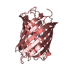
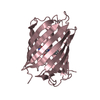
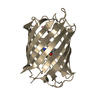
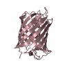
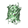
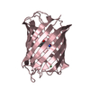
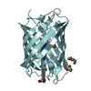
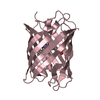
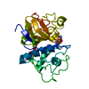
Fluorescent Protein mKeima at pH 3.8
Components
LARGE STOKES SHIFT FLUORESCENT PROTEIN
Keywords
FLUORESCENT PROTEIN / STOKES SHIFT / MKEIMA
Function / homology
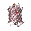
Green Fluorescent Protein / Green fluorescent protein / Green fluorescent protein-related / Green fluorescent protein / Green fluorescent protein / bioluminescence / Beta Barrel / Mainly Beta / Large stokes shift fluorescent protein
SHEET DETERMINATION METHOD: DSSP THE SHEETS PRESENTED AS "AA" IN EACH CHAIN ON SHEET RECORDS BELOW ... SHEET DETERMINATION METHOD: DSSP THE SHEETS PRESENTED AS "AA" IN EACH CHAIN ON SHEET RECORDS BELOW IS ACTUALLY AN 11-STRANDED BARREL THIS IS REPRESENTED BY A 12-STRANDED SHEET IN WHICH THE FIRST AND LAST STRANDS ARE IDENTICAL. THE SHEETS PRESENTED AS "BA" IN EACH CHAIN ON SHEET RECORDS BELOW IS ACTUALLY AN 11-STRANDED BARREL THIS IS REPRESENTED BY A 12-STRANDED SHEET IN WHICH THE FIRST AND LAST STRANDS ARE IDENTICAL. THE SHEETS PRESENTED AS "CA" IN EACH CHAIN ON SHEET RECORDS BELOW IS ACTUALLY AN 11-STRANDED BARREL THIS IS REPRESENTED BY A 12-STRANDED SHEET IN WHICH THE FIRST AND LAST STRANDS ARE IDENTICAL. THE SHEETS PRESENTED AS "DA" IN EACH CHAIN ON SHEET RECORDS BELOW IS ACTUALLY AN 11-STRANDED BARREL THIS IS REPRESENTED BY A 12-STRANDED SHEET IN WHICH THE FIRST AND LAST STRANDS ARE IDENTICAL.
A: LARGE STOKES SHIFT FLUORESCENT PROTEIN B: LARGE STOKES SHIFT FLUORESCENT PROTEIN C: LARGE STOKES SHIFT FLUORESCENT PROTEIN D: LARGE STOKES SHIFT FLUORESCENT PROTEIN hetero molecules
Resolution: 2.1→76.7 Å / Cor.coef. Fo:Fc: 0.959 / Cor.coef. Fo:Fc free: 0.927 / SU B: 5.128 / SU ML: 0.135 / Cross valid method: THROUGHOUT / ESU R: 0.207 / ESU R Free: 0.189 / Stereochemistry target values: MAXIMUM LIKELIHOOD Details: RESIDUE NUMEROTATION START AT MET 0 AS VAL 1 HAS BEEN INSERTED RESULTING FROM THE WT TO KOZAK CONSENSUS SEQUENCE MODIFICATION.
Rfactor
Num. reflection
% reflection
Selection details
Rfree
0.24788
3332
5.1 %
RANDOM
Rwork
0.19011
-
-
-
obs
0.19302
62294
99.23 %
-
Solvent computation
Ion probe radii: 0.8 Å / Shrinkage radii: 0.8 Å / VDW probe radii: 1.2 Å / Solvent model: MASK
 Movie
Movie Controller


 Open data
 Basic information
 Components
 Keywords
 Function and homology information
 Montipora sp. 20 (invertebrata)
 X-RAY DIFFRACTION /
  Authors
 Citation
 Structure visualization
 Downloads & links
 Other downloads


 PDBj
PDBj

 Assembly
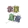
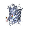


 Mass: 96.063 Da / Num. of mol.: 11 / Source method: obtained synthetically / Formula: SO4
 Mass: 18.015 Da / Num. of mol.: 761 / Source method: isolated from a natural source / Formula: H2O
 Sample preparation
 / Beamline: ID14-4 / Wavelength: 0.9395
  Processing