 Movie
Movie Controller

[English] 日本語
 Yorodumi
- PDB-1prz: Crystal structure of pseudouridine synthase RluD catalytic module -
+ Open data

- Basic information
| Entry | Database: PDB / ID: 1prz | ||||||
|---|---|---|---|---|---|---|---|
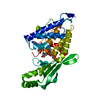
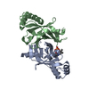
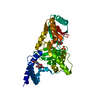
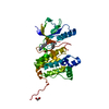
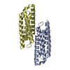
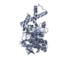
| Title | Crystal structure of pseudouridine synthase RluD catalytic module | ||||||
 Components Components | Ribosomal large subunit pseudouridine synthase D | ||||||
 Keywords Keywords | LYASE / Pseudouridine synthase / RluD / Montreal-Kingston Bacterial Structural Genomics Initiative / BSGI / Structural Genomics | ||||||
| Function / homology |  Function and homology information Function and homology information23S rRNA pseudouridine1911/1915/1917 synthase / 23S rRNA pseudouridine(1911/1915/1917) synthase activity / rRNA pseudouridine synthase activity / enzyme-directed rRNA pseudouridine synthesis / pseudouridine synthase activity / ribosomal large subunit assembly / rRNA binding / RNA binding / cytoplasm / cytosol Similarity search - Function | ||||||
| Biological species |  | ||||||
| Method |  X-RAY DIFFRACTION / MAD / Resolution: 1.8 Å X-RAY DIFFRACTION / MAD / Resolution: 1.8 Å | ||||||
 Authors Authors | Sivaraman, J. / Iannuzzi, P. / Cygler, M. / Matte, A. / Montreal-Kingston Bacterial Structural Genomics Initiative (BSGI) | ||||||
 Citation Citation | Journal: J.Mol.Biol. / Year: 2003 Title: Crystal structure of the RluD pseudouridine Synthase catalytic module, an enzyme that modifies 23S rRNA and is essential for normal cell growth of Escherichia coli Authors: Sivaraman, J. / Iannuzzi, P. / Cygler, M. / Matte, A. | ||||||
| History |
|
- Structure visualization
| Structure viewer | Molecule: MolmilJmol/JSmol |
|---|
- Downloads & links
-Download
| PDBx/mmCIF format | 1prz.cif.gz | 69.4 KB | Display | PDBx/mmCIF format |
|---|---|---|---|---|
| PDB format | pdb1prz.ent.gz | 50.6 KB | Display | PDB format |
| PDBx/mmJSON format | 1prz.json.gz | Tree view | PDBx/mmJSON format | |
| Others |  Other downloads Other downloads |
-Validation report
| Arichive directory | https://data.pdbj.org/pub/pdb/validation_reports/pr/1przftp://data.pdbj.org/pub/pdb/validation_reports/pr/1prz | HTTPS FTP |
|---|
-Related structure data
| Similar structure data | |
|---|---|
| Other databases |


-Links
 PDBj
PDBj- Assembly
| Deposited unit | 
| ||||||||||
|---|---|---|---|---|---|---|---|---|---|---|---|
| 1 |
| ||||||||||
| Unit cell |
|
-Components
| #1: Protein | Mass: 29229.068 Da / Num. of mol.: 1 / Fragment: catalytic domain Source method: isolated from a genetically manipulated source Source: (gene. exp.) References: UniProt: Q8X9F0, UniProt: P33643*PLUS, pseudouridylate synthase |
|---|---|
| #2: Water | ChemComp-HOH /  Mass: 18.015 Da / Num. of mol.: 294 / Source method: isolated from a natural source / Formula: H2O Mass: 18.015 Da / Num. of mol.: 294 / Source method: isolated from a natural source / Formula: H2O |
| Has protein modification | Y |
-Experimental details
-Experiment
| Experiment | Method: X-RAY DIFFRACTION / Number of used crystals: 1 |
|---|
- Sample preparation
| Crystal | Density Matthews: 2.64 Å3/Da / Density % sol: 53 % | |||||||||||||||||||||||||||||||||||||||||||||||||||||||||||||||
|---|---|---|---|---|---|---|---|---|---|---|---|---|---|---|---|---|---|---|---|---|---|---|---|---|---|---|---|---|---|---|---|---|---|---|---|---|---|---|---|---|---|---|---|---|---|---|---|---|---|---|---|---|---|---|---|---|---|---|---|---|---|---|---|---|
| Crystal grow | Temperature: 293 K / Method: microbatch under oil / pH: 7 Details: PEG 8000, KCL, Glycerol, pH 7., microbatch under oil, temperature 293K | |||||||||||||||||||||||||||||||||||||||||||||||||||||||||||||||
| Crystal grow | *PLUS Temperature: 4 ℃ / pH: 8 / Method: batch method | |||||||||||||||||||||||||||||||||||||||||||||||||||||||||||||||
| Components of the solutions | *PLUS
|
-Data collection
| Diffraction | Mean temperature: 100 K |
|---|---|
| Diffraction source | Source: ROTATING ANODE / Type: RIGAKU MICROMAX-007 / Wavelength: 1.5418 Å |
| Detector | Type: RIGAKU RAXIS HTC / Detector: IMAGE PLATE / Date: Feb 24, 2003 |
| Radiation | Monochromator: GRAPHITE / Protocol: MAD / Monochromatic (M) / Laue (L): M / Scattering type: x-ray |
| Radiation wavelength | Wavelength: 1.5418 Å / Relative weight: 1 |
| Reflection | Resolution: 1.8→50 Å / Num. all: 32444 / Num. obs: 32444 / % possible obs: 99.4 % / Observed criterion σ(I): -3 / Rsym value: 0.065 / Net I/σ(I): 12.4 |
| Reflection shell | Resolution: 1.8→1.86 Å / % possible all: 98.3 |
| Reflection | *PLUS Highest resolution: 1.8 Å / Num. measured all: 107285 / Rmerge(I) obs: 0.065 |
- Processing
| Software |
| |||||||||||||||||||||||||
|---|---|---|---|---|---|---|---|---|---|---|---|---|---|---|---|---|---|---|---|---|---|---|---|---|---|---|
| Refinement | Method to determine structure: MAD / Resolution: 1.8→50 Å / σ(F): 0 / σ(I): 0 / Stereochemistry target values: Engh & Huber
| |||||||||||||||||||||||||
| Refine analyze | Luzzati coordinate error obs: 0.29 Å | |||||||||||||||||||||||||
| Refinement step | Cycle: LAST / Resolution: 1.8→50 Å
| |||||||||||||||||||||||||
| Refine LS restraints |
| |||||||||||||||||||||||||
| Refinement | *PLUS Highest resolution: 1.8 Å / Lowest resolution: 45 Å | |||||||||||||||||||||||||
| Solvent computation | *PLUS | |||||||||||||||||||||||||
| Displacement parameters | *PLUS |
