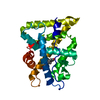
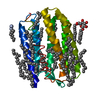
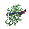
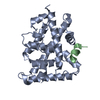
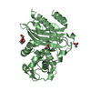
Entry Database : PDB / ID : 2v3kTitle The yeast ribosome synthesis factor Emg1 alpha beta knot fold methyltransferase ESSENTIAL FOR MITOTIC GROWTH 1 Keywords / / / / / / Function / homology Function Domain/homology Component
/ / / / / / / / / / / / / / / / / / / / / / / / / / / / / / / / / / / / Biological species SACCHAROMYCES CEREVISIAE (brewer's yeast)Method / / / Resolution : 2 Å Authors Leulliot, N. / Bohnsack, M.T. / Graille, M. / Tollervey, D. / VanTilbeurgh, H. Journal : Nucleic Acids Res. / Year : 2008Title : The Yeast Ribosome Synthesis Factor Emg1 is a Novel Member of the Superfamily of Alpha/Beta Knot Fold Methyltransferases.Authors : Leulliot, N. / Bohnsack, M.T. / Graille, M. / Tollervey, D. / Van Tilbeurgh, H. History Deposition Jun 18, 2007 Deposition site / Processing site Revision 1.0 Jul 10, 2007 Provider / Type Revision 1.1 May 8, 2011 Group Revision 1.2 Jul 13, 2011 Group Revision 1.3 Dec 13, 2023 Group Data collection / Database references ... Data collection / Database references / Derived calculations / Other / Refinement description Category chem_comp_atom / chem_comp_bond ... chem_comp_atom / chem_comp_bond / database_2 / pdbx_database_status / pdbx_initial_refinement_model / struct_conn Item _database_2.pdbx_DOI / _database_2.pdbx_database_accession ... _database_2.pdbx_DOI / _database_2.pdbx_database_accession / _pdbx_database_status.status_code_sf / _struct_conn.pdbx_leaving_atom_flag Revision 1.4 Oct 16, 2024 Group / Category / pdbx_modification_feature
Show all Show less
 Movie
Movie Controller

 Yorodumi
 Open data
 Basic information
 Components
 Keywords
 Function and homology information
 X-RAY DIFFRACTION /
  Authors
 Citation
 Structure visualization
 Downloads & links
 Other downloads
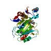
 PDBj
PDBj


 Assembly
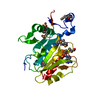


 Mass: 96.063 Da / Num. of mol.: 2 / Source method: obtained synthetically / Formula: SO4
 Mass: 398.437 Da / Num. of mol.: 1 / Source method: obtained synthetically / Formula: C15H22N6O5S
 Mass: 18.015 Da / Num. of mol.: 141 / Source method: isolated from a natural source / Formula: H2O
 Sample preparation
 / Beamline: ID14-4 / Wavelength: 0.931
  Processing