- PDB-5c7h: Crystal structure of aldo-keto reductase from Sinorhizobium melil... -
+
Open data
ID or keywords:
Loading...
-
Basic information
Entry
Database: PDB / ID: 5c7h
Title
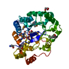
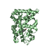
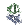
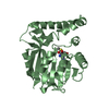
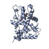
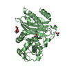
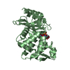
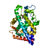
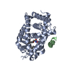
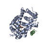
Crystal structure of aldo-keto reductase from Sinorhizobium meliloti 1021 in complex with NADPH
Components
Aldo-keto reductase
Keywords
OXIDOREDUCTASE / aldo-keto reductase / AKR / NADPH / NYSGRC / PSI-Biology / Structural Genomics / New York Structural Genomics Research Consortium
Function / homology
Function and homology information
Oxidoreductases; Acting on the CH-OH group of donors; With NAD+ or NADP+ as acceptor / oxidoreductase activity / nucleotide binding Similarity search - Function
Mass: 30757.080 Da / Num. of mol.: 1 Source method: isolated from a genetically manipulated source Source: (gene. exp.) Rhizobium meliloti (strain 1021) (bacteria) Strain: 1021 / Gene: SMc01429 / Plasmid: pSGC-His / Production host: Escherichia coli (E. coli) / Strain (production host): BL21 (DE3) RIL References: UniProt: Q92NR7, Oxidoreductases; Acting on the CH-OH group of donors; With NAD+ or NADP+ as acceptor
Mass: 18.015 Da / Num. of mol.: 417 / Source method: isolated from a natural source / Formula: H2O
-
Experimental details
-
Experiment
Experiment
Method: X-RAY DIFFRACTION / Number of used crystals: 1
-
Sample preparation
Crystal
Density Matthews: 2.24 Å3/Da / Density % sol: 45.1 %
Crystal grow
Temperature: 289 K / Method: vapor diffusion, hanging drop / pH: 7.5 Details: 0.2 ul of 14 mg/ml protein in 20 mM HEPES pH 7.5, 150 mM NaCl, 10% Glycerol, 0.1% Sodium Azide and 0.5 mM TCEP were mixed with 0.2 ul of the MCSG 2 condition #28 (0.2 M Ammonium Citrate ...Details: 0.2 ul of 14 mg/ml protein in 20 mM HEPES pH 7.5, 150 mM NaCl, 10% Glycerol, 0.1% Sodium Azide and 0.5 mM TCEP were mixed with 0.2 ul of the MCSG 2 condition #28 (0.2 M Ammonium Citrate Tribasic pH 7.0, 20% (w/v) PEG 3350) and equilibrated against 1.5 M NaCl solution in 96 Well 3 drop Crystallization Plate (Swissci). Before crystallization protein was incubated with 1/50 v/v of 1 mg/ml TEV solution at 289 K for 1 hour PH range: 7.0-7.5
In the structure databanks used in Yorodumi, some data are registered as the other names, "COVID-19 virus" and "2019-nCoV". Here are the details of the virus and the list of structure data.
Jan 31, 2019. EMDB accession codes are about to change! (news from PDBe EMDB page)
EMDB accession codes are about to change! (news from PDBe EMDB page)
The allocation of 4 digits for EMDB accession codes will soon come to an end. Whilst these codes will remain in use, new EMDB accession codes will include an additional digit and will expand incrementally as the available range of codes is exhausted. The current 4-digit format prefixed with “EMD-” (i.e. EMD-XXXX) will advance to a 5-digit format (i.e. EMD-XXXXX), and so on. It is currently estimated that the 4-digit codes will be depleted around Spring 2019, at which point the 5-digit format will come into force.
The EM Navigator/Yorodumi systems omit the EMD- prefix.
Related info.:Q: What is EMD? / ID/Accession-code notation in Yorodumi/EM Navigator
Yorodumi is a browser for structure data from EMDB, PDB, SASBDB, etc.
This page is also the successor to EM Navigator detail page, and also detail information page/front-end page for Omokage search.
The word "yorodu" (or yorozu) is an old Japanese word meaning "ten thousand". "mi" (miru) is to see.
Related info.:EMDB / PDB / SASBDB / Comparison of 3 databanks / Yorodumi Search / Aug 31, 2016. New EM Navigator & Yorodumi / Yorodumi Papers / Jmol/JSmol / Function and homology information / Changes in new EM Navigator and Yorodumi
 Movie
Movie Controller

 Yorodumi
 Open data
 Basic information
 Components
 Keywords
 Function and homology information
 Rhizobium meliloti (bacteria)
 X-RAY DIFFRACTION /
  Authors
 United States, 1items
  Citation
 Structure visualization
 Downloads & links
 Other downloads
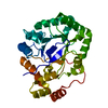
 PDBj
PDBj


 Assembly
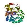

 Mass: 745.421 Da / Num. of mol.: 1 / Source method: obtained synthetically / Formula: C21H30N7O17P3
 Mass: 18.015 Da / Num. of mol.: 417 / Source method: isolated from a natural source / Formula: H2O
 Sample preparation
 Processing