- PDB-4pmj: Crystal structure of a putative oxidoreductase from Sinorhizobium... -
+
Open data
ID or keywords:
Loading...
-
Basic information
Entry
Database: PDB / ID: 4pmj
Title
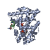
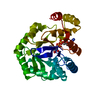
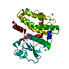
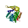
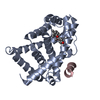
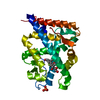
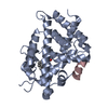
Crystal structure of a putative oxidoreductase from Sinorhizobium meliloti 1021 in complex with NADP
Components
Putative oxidoreductase
Keywords
OXIDOREDUCTASE / Structural Genomics / PSI-Biology / New York Structural Genomics Research Consortium / NYSGRC / putative oxidoreductase / NADP / limited proteolysis.
Function / homology
Function and homology information
Oxidoreductases; Acting on the CH-OH group of donors; With NAD+ or NADP+ as acceptor / oxidoreductase activity / nucleotide binding Similarity search - Function
Mass: 31085.338 Da / Num. of mol.: 1 Source method: isolated from a genetically manipulated source Source: (gene. exp.) Rhizobium meliloti (bacteria) / Strain: 1021 / Gene: R02112, SMc01429 / Plasmid: pSGC-His / Production host: Escherichia coli (E. coli) / Strain (production host): BL21 (DE3) RIL References: UniProt: Q92NR7, Oxidoreductases; Acting on the CH-OH group of donors; With NAD+ or NADP+ as acceptor
Mass: 18.015 Da / Num. of mol.: 115 / Source method: isolated from a natural source / Formula: H2O
Has protein modification
Y
Sequence details
The protein was expressed with His tag and subjected to limited proteolysis by chymotrypsin
-
Experimental details
-
Experiment
Experiment
Method: X-RAY DIFFRACTION
-
Sample preparation
Crystal
Density Matthews: 2.26 Å3/Da / Density % sol: 45.61 %
Crystal grow
Temperature: 289 K / Method: vapor diffusion, sitting drop / pH: 7 Details: 0.2 ul of 14 mg/ml protein in 20 mM HEPES pH 7.5, 150 mM NaCl, 10% Glycerol, 0.1% Sodium Azide and 0.5 mM TCEP were mixed with 0.2 ul of the MCSG-II condition #42 (1.1 M Ammonium Tartrate ...Details: 0.2 ul of 14 mg/ml protein in 20 mM HEPES pH 7.5, 150 mM NaCl, 10% Glycerol, 0.1% Sodium Azide and 0.5 mM TCEP were mixed with 0.2 ul of the MCSG-II condition #42 (1.1 M Ammonium Tartrate Dibasic pH 7.0) and equilibrated against 1.5 M NaCl solution in 96 Well 3 drop Crystallization Plate (Swissci). Before crystallization protein was incubated with 1/50 v/v of 2 mg/ml chymotrypsin solution at 289 K for 3 hours. Temp details: Rigaku Gallery 700
Resolution: 2.2→2.24 Å / Redundancy: 4.1 % / Rmerge(I) obs: 0.457 / Mean I/σ(I) obs: 1.9 / % possible all: 85.7
-
Processing
Software
Name
Version
Classification
HKL-3000
datacollection
HKL-3000
phasing
HKL-3000
datascaling
SHELXDE
phasing
MLPHARE
phasing
DM
phasing
Coot
modelbuilding
REFMAC
5.8.0049
refinement
PDB_EXTRACT
dataextraction
Refinement
Method to determine structure: SAD / Resolution: 2.2→50 Å / Cor.coef. Fo:Fc: 0.955 / Cor.coef. Fo:Fc free: 0.93 / SU B: 12.06 / SU ML: 0.162 / SU R Cruickshank DPI: 0.15 / Cross valid method: THROUGHOUT / σ(F): 0 / ESU R: 0.271 / ESU R Free: 0.201 / SU Rfree Cruickshank DPI: 0.2 Stereochemistry target values: MAXIMUM LIKELIHOOD WITH PHASES Details: HYDROGENS HAVE BEEN ADDED IN THE RIDING POSITIONS
Rfactor
Num. reflection
% reflection
Selection details
Rfree
0.21676
731
5.1 %
RANDOM
Rwork
0.16725
-
-
-
obs
0.16977
13677
97.87 %
-
Solvent computation
Ion probe radii: 0.8 Å / Shrinkage radii: 0.8 Å / VDW probe radii: 1.2 Å / Solvent model: MASK
 Movie
Movie Controller

 Yorodumi
 Open data
 Basic information
 Components
 Keywords
 Function and homology information
 Rhizobium meliloti (bacteria)
 X-RAY DIFFRACTION /
  Authors
 United States, 1items
  Citation
 Structure visualization
 Downloads & links
 Other downloads
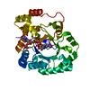


 PDBj
PDBj
 Assembly
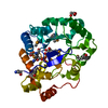

 Mass: 743.405 Da / Num. of mol.: 1 / Source method: obtained synthetically / Formula: C21H28N7O17P3
 Mass: 92.094 Da / Num. of mol.: 1 / Source method: obtained synthetically / Formula: C3H8O3
 Mass: 18.015 Da / Num. of mol.: 115 / Source method: isolated from a natural source / Formula: H2O
 Sample preparation
 Processing