 Movie
Movie Controller

[English] 日本語
 Yorodumi
- PDB-3t7t: Crystal structure of complex of SAH and BVU_3255, a methyltransfe... -
+ Open data

- Basic information
| Entry | Database: PDB / ID: 3t7t | ||||||
|---|---|---|---|---|---|---|---|
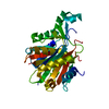
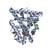
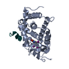
| Title | Crystal structure of complex of SAH and BVU_3255, a methyltransferase from Bacteroides vulgatus ATCC 8482 | ||||||
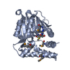
 Components Components | Putative methyltransferase | ||||||
 Keywords Keywords | TRANSFERASE / Small molecule methyltransferase / BVU_3255 / ROSSMANN FOLD / METHYLTRASNFERASE / SAM / Methylation | ||||||
| Function / homology |  Function and homology information Function and homology information | ||||||
| Biological species |  Bacteroides vulgatus (bacteria) Bacteroides vulgatus (bacteria) | ||||||
| Method |  X-RAY DIFFRACTION / MOLECULAR REPLACEMENT / Resolution: 2.5 Å X-RAY DIFFRACTION / MOLECULAR REPLACEMENT / Resolution: 2.5 Å | ||||||
 Authors Authors | Kumar, V. / Sivaraman, J. | ||||||
 Citation Citation | Journal: J.Struct.Biol. / Year: 2011 Title: Structural characterization of BVU_3255, a methyltransferase from human intestine antibiotic resistant pathogen Bacteroides vulgatus Authors: Kumar, V. / Sivaraman, J. | ||||||
| History |
|
- Structure visualization
| Structure viewer | Molecule: MolmilJmol/JSmol |
|---|
- Downloads & links
-Download
| PDBx/mmCIF format | 3t7t.cif.gz | 208.8 KB | Display | PDBx/mmCIF format |
|---|---|---|---|---|
| PDB format | pdb3t7t.ent.gz | 166.4 KB | Display | PDB format |
| PDBx/mmJSON format | 3t7t.json.gz | Tree view | PDBx/mmJSON format | |
| Others |  Other downloads Other downloads |
-Validation report
| Arichive directory | https://data.pdbj.org/pub/pdb/validation_reports/t7/3t7tftp://data.pdbj.org/pub/pdb/validation_reports/t7/3t7t | HTTPS FTP |
|---|
-Related structure data
| Related structure data |  3t7rC  3t7sC  3e7pS C: citing same article ( S: Starting model for refinement |
|---|---|
| Similar structure data |


-Links
 PDBj
PDBj- Assembly
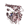
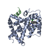
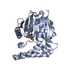
| Deposited unit | 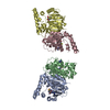
| ||||||||
|---|---|---|---|---|---|---|---|---|---|
| 1 | 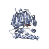
| ||||||||
| 2 | 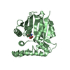
| ||||||||
| 3 | 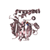
| ||||||||
| 4 | 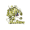
| ||||||||
| Unit cell |
|
-Components
| #1: Protein | Mass: 30664.715 Da / Num. of mol.: 4 Source method: isolated from a genetically manipulated source Source: (gene. exp.) Bacteroides vulgatus (bacteria) / Strain: ATCC 8482 / Gene: BVU_3255 / Plasmid: pGS21a / Production host: #2: Chemical | ChemComp-SAH /   Type: L-peptide linking / Mass: 384.411 Da / Num. of mol.: 4 / Source method: obtained synthetically / Formula: C14H20N6O5S Type: L-peptide linking / Mass: 384.411 Da / Num. of mol.: 4 / Source method: obtained synthetically / Formula: C14H20N6O5S#3: Water | ChemComp-HOH / |  Mass: 18.015 Da / Num. of mol.: 287 / Source method: isolated from a natural source / Formula: H2O Mass: 18.015 Da / Num. of mol.: 287 / Source method: isolated from a natural source / Formula: H2O |
|---|
-Experimental details
-Experiment
| Experiment | Method: X-RAY DIFFRACTION / Number of used crystals: 1 |
|---|
- Sample preparation
| Crystal | Density Matthews: 2.15 Å3/Da / Density % sol: 42.75 % |
|---|---|
| Crystal grow | Temperature: 298 K / Method: vapor diffusion, hanging drop Details: 120mM (NH4)(NO3), 16% w/v PEG 3350, 200mM non-detergent sulfobetaine 221 (NDSB-221), VAPOR DIFFUSION, HANGING DROP, temperature 298K |
-Data collection
| Diffraction | Mean temperature: 100 K |
|---|---|
| Diffraction source | Source: ROTATING ANODE / Type: BRUKER AXS MICROSTAR / Wavelength: 1.5418 Å |
| Detector | Type: Bruker Platinum 135 / Detector: CCD / Date: Nov 30, 2010 |
| Radiation | Monochromator: Graphite / Protocol: SINGLE WAVELENGTH / Monochromatic (M) / Laue (L): M / Scattering type: x-ray |
| Radiation wavelength | Wavelength: 1.5418 Å / Relative weight: 1 |
| Reflection | Resolution: 2.5→50 Å / Num. all: 35718 / Num. obs: 35718 / % possible obs: 100 % / Observed criterion σ(F): 1 / Observed criterion σ(I): 1 / Rsym value: 0.072 / Net I/σ(I): 10.42 |
- Processing
| Software |
| ||||||||||||||||||||||||||||
|---|---|---|---|---|---|---|---|---|---|---|---|---|---|---|---|---|---|---|---|---|---|---|---|---|---|---|---|---|---|
| Refinement | Method to determine structure: MOLECULAR REPLACEMENT Starting model: PDB ENTRY 3E7P Resolution: 2.5→30 Å / Occupancy max: 1 / Occupancy min: 1 / Isotropic thermal model: Isotropic / Cross valid method: THROUGHOUT / σ(F): 3 / Stereochemistry target values: Engh & Huber
| ||||||||||||||||||||||||||||
| Solvent computation | Bsol: 24.4665 Å2 | ||||||||||||||||||||||||||||
| Displacement parameters | Biso max: 84.82 Å2 / Biso mean: 31.8601 Å2 / Biso min: 4.71 Å2
| ||||||||||||||||||||||||||||
| Refinement step | Cycle: LAST / Resolution: 2.5→30 Å
| ||||||||||||||||||||||||||||
| Refine LS restraints |
| ||||||||||||||||||||||||||||
| Xplor file |
|
