 Movie
Movie Controller


+ Open data

- Basic information
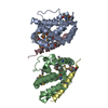
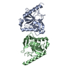
| Entry | Database: PDB / ID: 2uzq | ||||||
|---|---|---|---|---|---|---|---|
| Title | Protein Phosphatase, New Crystal Form | ||||||
 Components Components | M-PHASE INDUCER PHOSPHATASE 2 | ||||||
 Keywords Keywords | HYDROLASE / CELL DIVISION / PHOSPHORYLATION / DUAL SPECIFICITY / MITOSIS / CELL CYCLE / PHOSPHATASE / PROTEIN PHOSPHATASE | ||||||
| Function / homology |  Function and homology information Function and homology informationpositive regulation of G2/MI transition of meiotic cell cycle / oocyte maturation / female meiosis I / Deregulated CDK5 triggers multiple neurodegenerative pathways in Alzheimer's disease models / positive regulation of cytokinesis / positive regulation of G2/M transition of mitotic cell cycle / Regulation of MITF-M-dependent genes involved in cell cycle and proliferation / Cyclin A:Cdk2-associated events at S phase entry / Cyclin A/B1/B2 associated events during G2/M transition / phosphoprotein phosphatase activity ...positive regulation of G2/MI transition of meiotic cell cycle / oocyte maturation / female meiosis I / Deregulated CDK5 triggers multiple neurodegenerative pathways in Alzheimer's disease models / positive regulation of cytokinesis / positive regulation of G2/M transition of mitotic cell cycle / Regulation of MITF-M-dependent genes involved in cell cycle and proliferation / Cyclin A:Cdk2-associated events at S phase entry / Cyclin A/B1/B2 associated events during G2/M transition / phosphoprotein phosphatase activity / protein-tyrosine-phosphatase / positive regulation of mitotic cell cycle / protein tyrosine phosphatase activity / G2/M transition of mitotic cell cycle / spindle pole / mitotic cell cycle / protein phosphorylation / cell division / positive regulation of cell population proliferation / centrosome / protein kinase binding / nucleoplasm / nucleus / cytoplasm / cytosol Similarity search - Function | ||||||
| Biological species |  HOMO SAPIENS (human) HOMO SAPIENS (human) | ||||||
| Method |  X-RAY DIFFRACTION / SYNCHROTRON / MOLECULAR REPLACEMENT / Resolution: 2.38 Å X-RAY DIFFRACTION / SYNCHROTRON / MOLECULAR REPLACEMENT / Resolution: 2.38 Å | ||||||
 Authors Authors | Hillig, R.C. / Eberspaecher, U. | ||||||
 Citation Citation | Journal: To be Published Title: New Crystal Form of Protein Phosphatase Cdc25B Triggered by Guanidinium Chloride as an Additive Authors: Hillig, R.C. / Eberspaecher, U. #1: Journal: Biochemistry / Year: 2005Title: Experimental Validation of the Docking Orientation of Cdc25 with its Cdk2-Cyca Protein Substrate. Authors: Sohn, J. / Parks, J.M. / Buhrman, G. / Brown, P. / Kristjansdottir, K. / Safi, A. / Edelsbrunner, H. / Yang, W. / Rudolph, J. #2: Journal: J.Mol.Biol. / Year: 1999Title: Crystal Structure of the Catalytic Subunit of Cdc25B Required for G2/M Phase Transition of the Cell Cycle. Authors: Reynolds, R.A. / Yem, A.W. / Wolfe, C.L. / Deibel, M.R.J. / Chidester, C.G. / Watenpaugh, K.D. | ||||||
| History |
|
- Structure visualization
| Structure viewer | Molecule: MolmilJmol/JSmol |
|---|
- Downloads & links
-Download
| PDBx/mmCIF format | 2uzq.cif.gz | 222.5 KB | Display | PDBx/mmCIF format |
|---|---|---|---|---|
| PDB format | pdb2uzq.ent.gz | 180 KB | Display | PDB format |
| PDBx/mmJSON format | 2uzq.json.gz | Tree view | PDBx/mmJSON format | |
| Others |  Other downloads Other downloads |
-Validation report
| Arichive directory | https://data.pdbj.org/pub/pdb/validation_reports/uz/2uzqftp://data.pdbj.org/pub/pdb/validation_reports/uz/2uzq | HTTPS FTP |
|---|
-Related structure data
| Related structure data |  1qb0S S: Starting model for refinement |
|---|---|
| Similar structure data |


-Links
 PDBj
PDBj


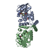
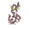
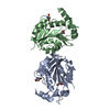
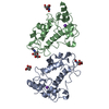
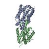
- Assembly
| Deposited unit | 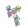
| ||||||||||||||||||||||||
|---|---|---|---|---|---|---|---|---|---|---|---|---|---|---|---|---|---|---|---|---|---|---|---|---|---|
| 1 | 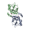
| ||||||||||||||||||||||||
| 2 | 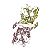
| ||||||||||||||||||||||||
| 3 | 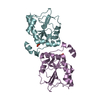
| ||||||||||||||||||||||||
| Unit cell |
| ||||||||||||||||||||||||
| Noncrystallographic symmetry (NCS) | NCS oper:
|
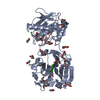
-Components
| #1: Protein | Mass: 23450.812 Da / Num. of mol.: 6 / Fragment: CATALYTIC DOMAIN, RESIDUES 377-566 Source method: isolated from a genetically manipulated source Source: (gene. exp.) HOMO SAPIENS (human) / Production host:  #2: Chemical | ChemComp-PO4 /   Mass: 94.971 Da / Num. of mol.: 6 / Source method: obtained synthetically / Formula: PO4 Mass: 94.971 Da / Num. of mol.: 6 / Source method: obtained synthetically / Formula: PO4#3: Water | ChemComp-HOH / |  Mass: 18.015 Da / Num. of mol.: 196 / Source method: isolated from a natural source / Formula: H2O Mass: 18.015 Da / Num. of mol.: 196 / Source method: isolated from a natural source / Formula: H2OSequence details | SEQUENCE CONTAINS N-TERMINAL RESIDUES WHICH REPRESENT A CLONING ARTIFACT (THROMBIN CLEAVAGE SITE ...SEQUENCE CONTAINS N-TERMINAL RESIDUES WHICH REPRESENT A CLONING ARTIFACT (THROMBIN CLEAVAGE SITE AND POLY GLY LINKER, GSPGIS GGGGG). OUT OF THE FOUR ISOFORM SEQUENCES GENERATED BY ALTERNATE SPLICING, THE ENTRY BELONGS TO ISOFORM 1 | |
|---|
-Experimental details
-Experiment
| Experiment | Method: X-RAY DIFFRACTION / Number of used crystals: 1 |
|---|
- Sample preparation
| Crystal | Density Matthews: 2.7 Å3/Da / Density % sol: 53.4 % |
|---|---|
| Crystal grow | pH: 7 / Details: PEG 8000, HEPES PH 7.0, GUANIDINIUM CHLORIDE |
-Data collection
| Diffraction | Mean temperature: 100 K |
|---|---|
| Diffraction source | Source: SYNCHROTRON / Site: BESSY  / Beamline: 14.1 / Wavelength: 0.95368 / Beamline: 14.1 / Wavelength: 0.95368 |
| Detector | Type: MARRESEARCH / Detector: CCD / Date: Sep 30, 2002 |
| Radiation | Protocol: SINGLE WAVELENGTH / Monochromatic (M) / Laue (L): M / Scattering type: x-ray |
| Radiation wavelength | Wavelength: 0.95368 Å / Relative weight: 1 |
| Reflection | Resolution: 2.38→36.8 Å / Num. obs: 58835 / % possible obs: 97.3 % / Observed criterion σ(I): 0 / Redundancy: 2.9 % / Biso Wilson estimate: 41.5 Å2 / Rmerge(I) obs: 0.05 |
| Reflection shell | Resolution: 2.38→2.47 Å / Redundancy: 2.5 % / Rmerge(I) obs: 0.33 / Mean I/σ(I) obs: 2.5 / % possible all: 89.3 |
- Processing
| Software |
| ||||||||||||||||||||||||||||||||||||||||||||||||||||||||||||||||||||||||||||||||
|---|---|---|---|---|---|---|---|---|---|---|---|---|---|---|---|---|---|---|---|---|---|---|---|---|---|---|---|---|---|---|---|---|---|---|---|---|---|---|---|---|---|---|---|---|---|---|---|---|---|---|---|---|---|---|---|---|---|---|---|---|---|---|---|---|---|---|---|---|---|---|---|---|---|---|---|---|---|---|---|---|---|
| Refinement | Method to determine structure: MOLECULAR REPLACEMENT Starting model: PDB ENTRY 1QB0 Resolution: 2.38→36.77 Å / Rfactor Rfree error: 0.004 / Isotropic thermal model: RESTRAINED / Cross valid method: THROUGHOUT / σ(F): 0
| ||||||||||||||||||||||||||||||||||||||||||||||||||||||||||||||||||||||||||||||||
| Solvent computation | Solvent model: FLAT MODEL / Bsol: 37.1889 Å2 / ksol: 0.355341 e/Å3 | ||||||||||||||||||||||||||||||||||||||||||||||||||||||||||||||||||||||||||||||||
| Displacement parameters | Biso mean: 54 Å2
| ||||||||||||||||||||||||||||||||||||||||||||||||||||||||||||||||||||||||||||||||
| Refine analyze |
| ||||||||||||||||||||||||||||||||||||||||||||||||||||||||||||||||||||||||||||||||
| Refinement step | Cycle: LAST / Resolution: 2.38→36.77 Å
| ||||||||||||||||||||||||||||||||||||||||||||||||||||||||||||||||||||||||||||||||
| Refine LS restraints |
| ||||||||||||||||||||||||||||||||||||||||||||||||||||||||||||||||||||||||||||||||
| Refine LS restraints NCS | NCS model details: NCS RESTRAINTS | ||||||||||||||||||||||||||||||||||||||||||||||||||||||||||||||||||||||||||||||||
| LS refinement shell | Resolution: 2.38→2.47 Å / Rfactor Rfree error: 0.021 / Total num. of bins used: 10 /
| ||||||||||||||||||||||||||||||||||||||||||||||||||||||||||||||||||||||||||||||||
| Xplor file | Serial no: 1 / Param file: PROTEIN_REP.PARAM / Topol file: ION.TOP |
