 Movie
Movie Controller

[English] 日本語
 Yorodumi
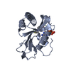
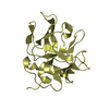
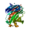
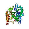
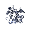
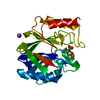
- PDB-2wee: Crystal structure of Rv0371c from Mycobacterium tuberculosis H37Rv -
+ Open data

- Basic information
| Entry | Database: PDB / ID: 2wee | ||||||
|---|---|---|---|---|---|---|---|
| Title | Crystal structure of Rv0371c from Mycobacterium tuberculosis H37Rv | ||||||
 Components Components | MOBA-RELATED PROTEIN | ||||||
 Keywords Keywords | UNKNOWN FUNCTION | ||||||
| Function / homology |  Function and homology information Function and homology information | ||||||
| Biological species |  MYCOBACTERIUM TUBERCULOSIS H37RV (bacteria) MYCOBACTERIUM TUBERCULOSIS H37RV (bacteria) | ||||||
| Method |  X-RAY DIFFRACTION / SYNCHROTRON / MOLECULAR REPLACEMENT / Resolution: 1.65 Å X-RAY DIFFRACTION / SYNCHROTRON / MOLECULAR REPLACEMENT / Resolution: 1.65 Å | ||||||
 Authors Authors | Cho, H.J. / Kang, B.S. | ||||||
 Citation Citation | Journal: To be Published Title: Crystal Structure of Rv0371C from Mycobacterium Tuberculosis H37Rv Authors: Cho, H.J. / Kang, B.S. | ||||||
| History |
|
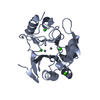
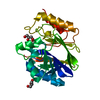
- Structure visualization
| Structure viewer | Molecule: MolmilJmol/JSmol |
|---|
- Downloads & links
-Download
| PDBx/mmCIF format | 2wee.cif.gz | 93.3 KB | Display | PDBx/mmCIF format |
|---|---|---|---|---|
| PDB format | pdb2wee.ent.gz | 72 KB | Display | PDB format |
| PDBx/mmJSON format | 2wee.json.gz | Tree view | PDBx/mmJSON format | |
| Others |  Other downloads Other downloads |
-Validation report
| Arichive directory | https://data.pdbj.org/pub/pdb/validation_reports/we/2weeftp://data.pdbj.org/pub/pdb/validation_reports/we/2wee | HTTPS FTP |
|---|
-Related structure data
| Related structure data | 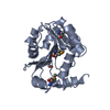 2we9SC S: Starting model for refinement C: citing same article ( |
|---|---|
| Similar structure data |

-Links
 PDBj
PDBj

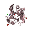
- Assembly
| Deposited unit | 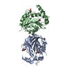
| ||||||||
|---|---|---|---|---|---|---|---|---|---|
| 1 | 
| ||||||||
| 2 | 
| ||||||||
| Unit cell |
|
-Components
| #1: Protein | Mass: 21158.334 Da / Num. of mol.: 2 / Source method: isolated from a natural source Source: (natural) MYCOBACTERIUM TUBERCULOSIS H37RV (bacteria)References: UniProt: O53706 #2: Chemical |   Mass: 92.094 Da / Num. of mol.: 2 / Source method: obtained synthetically / Formula: C3H8O3 Mass: 92.094 Da / Num. of mol.: 2 / Source method: obtained synthetically / Formula: C3H8O3#3: Water | ChemComp-HOH / |  Mass: 18.015 Da / Num. of mol.: 338 / Source method: isolated from a natural source / Formula: H2O Mass: 18.015 Da / Num. of mol.: 338 / Source method: isolated from a natural source / Formula: H2OHas protein modification | Y | |
|---|
-Experimental details
-Experiment
| Experiment | Method: X-RAY DIFFRACTION / Number of used crystals: 1 |
|---|
- Sample preparation
| Crystal | Density Matthews: 2.2 Å3/Da / Density % sol: 44 % / Description: NONE |
|---|---|
| Crystal grow | pH: 7.5 / Details: 0.1 M MES PH 6.0 AND 18% PEG 4000 BY MICRO-SEEDING |
-Data collection
| Diffraction | Mean temperature: 294 K |
|---|---|
| Diffraction source | Source: SYNCHROTRON / Site: PAL/PLS  / Beamline: 6C1 / Wavelength: 1 / Beamline: 6C1 / Wavelength: 1 |
| Detector | Type: ADSC CCD / Detector: CCD / Date: Nov 12, 2008 / Details: MIRRORS |
| Radiation | Protocol: SINGLE WAVELENGTH / Monochromatic (M) / Laue (L): M / Scattering type: x-ray |
| Radiation wavelength | Wavelength: 1 Å / Relative weight: 1 |
| Reflection | Resolution: 1.65→50 Å / Num. obs: 40721 / % possible obs: 89.4 % / Observed criterion σ(I): 1.65 / Redundancy: 7.1 % / Rmerge(I) obs: 0.05 / Net I/σ(I): 37.6 |
| Reflection shell | Resolution: 1.65→1.71 Å / Redundancy: 6.9 % / Rmerge(I) obs: 0.49 / Mean I/σ(I) obs: 3.36 / % possible all: 98.3 |
- Processing
| Software |
| ||||||||||||||||||||||||||||||||||||||||||||||||||||||||||||||||||||||||||||||||||||||||||||||||||||||||||||||||||||||||||||||||||||||||||||||||||||||||||||||||||||||||||||||||||||||
|---|---|---|---|---|---|---|---|---|---|---|---|---|---|---|---|---|---|---|---|---|---|---|---|---|---|---|---|---|---|---|---|---|---|---|---|---|---|---|---|---|---|---|---|---|---|---|---|---|---|---|---|---|---|---|---|---|---|---|---|---|---|---|---|---|---|---|---|---|---|---|---|---|---|---|---|---|---|---|---|---|---|---|---|---|---|---|---|---|---|---|---|---|---|---|---|---|---|---|---|---|---|---|---|---|---|---|---|---|---|---|---|---|---|---|---|---|---|---|---|---|---|---|---|---|---|---|---|---|---|---|---|---|---|---|---|---|---|---|---|---|---|---|---|---|---|---|---|---|---|---|---|---|---|---|---|---|---|---|---|---|---|---|---|---|---|---|---|---|---|---|---|---|---|---|---|---|---|---|---|---|---|---|---|
| Refinement | Method to determine structure: MOLECULAR REPLACEMENT Starting model: PDB ENTRY 2WE9 Resolution: 1.65→31.25 Å / Cor.coef. Fo:Fc: 0.957 / Cor.coef. Fo:Fc free: 0.926 / SU B: 6.168 / SU ML: 0.104 / TLS residual ADP flag: LIKELY RESIDUAL / Cross valid method: THROUGHOUT / ESU R: 0.134 / ESU R Free: 0.136 / Stereochemistry target values: MAXIMUM LIKELIHOOD / Details: HYDROGENS HAVE BEEN ADDED IN THE RIDING POSITIONS.
| ||||||||||||||||||||||||||||||||||||||||||||||||||||||||||||||||||||||||||||||||||||||||||||||||||||||||||||||||||||||||||||||||||||||||||||||||||||||||||||||||||||||||||||||||||||||
| Solvent computation | Ion probe radii: 0.8 Å / Shrinkage radii: 0.8 Å / VDW probe radii: 1.2 Å / Solvent model: MASK | ||||||||||||||||||||||||||||||||||||||||||||||||||||||||||||||||||||||||||||||||||||||||||||||||||||||||||||||||||||||||||||||||||||||||||||||||||||||||||||||||||||||||||||||||||||||
| Displacement parameters | Biso mean: 21.68 Å2
| ||||||||||||||||||||||||||||||||||||||||||||||||||||||||||||||||||||||||||||||||||||||||||||||||||||||||||||||||||||||||||||||||||||||||||||||||||||||||||||||||||||||||||||||||||||||
| Refinement step | Cycle: LAST / Resolution: 1.65→31.25 Å
| ||||||||||||||||||||||||||||||||||||||||||||||||||||||||||||||||||||||||||||||||||||||||||||||||||||||||||||||||||||||||||||||||||||||||||||||||||||||||||||||||||||||||||||||||||||||
| Refine LS restraints |
|