Protocol: SINGLE WAVELENGTH / Monochromatic (M) / Laue (L): M / Scattering type: x-ray
Radiation wavelength
Wavelength: 0.97853 Å / Relative weight: 1
Reflection twin
Crystal-ID
ID
Operator
Domain-ID
Fraction
1
1
H, K, L
1
0.631
1
1
K, H, -L
2
0.369
Reflection
Resolution: 2.2→50 Å / Num. obs: 52682 / % possible obs: 100 % / Redundancy: 10.3 % / Net I/σ(I): 36.78
-
Processing
Software
Name
Version
Classification
REFMAC
5.8.0103
refinement
SCALEPACK
datareduction
HKL-2000
datascaling
PHASER
phasing
Refinement
Resolution: 2.2→31.91 Å / Cor.coef. Fo:Fc: 0.921 / Cor.coef. Fo:Fc free: 0.865 / SU B: 8.283 / SU ML: 0.219 / Cross valid method: THROUGHOUT / ESU R: 0.077 / ESU R Free: 0.055 / Stereochemistry target values: MAXIMUM LIKELIHOOD / Details: HYDROGENS HAVE BEEN ADDED IN THE RIDING POSITIONS
Rfactor
Num. reflection
% reflection
Selection details
Rfree
0.27733
2482
4.9 %
RANDOM
Rwork
0.21411
-
-
-
obs
0.21722
47813
95.5 %
-
Solvent computation
Ion probe radii: 0.8 Å / Shrinkage radii: 0.8 Å / VDW probe radii: 1.2 Å / Solvent model: MASK
 Movie
Movie Controller

 Yorodumi
 Open data
 Basic information
 Components
 Keywords
 Function and homology information
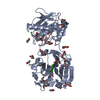
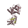
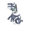
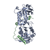
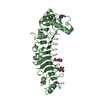
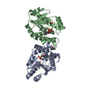
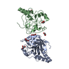
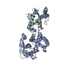
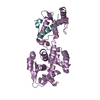
 Lake Victoria marburgvirus
 X-RAY DIFFRACTION /
  Authors
 Citation
 Structure visualization
 Downloads & links
 Other downloads

 PDBj
PDBj
 Assembly
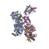


 Mass: 96.063 Da / Num. of mol.: 3 / Source method: isolated from a natural source / Formula: SO4
 Mass: 18.015 Da / Num. of mol.: 232 / Source method: isolated from a natural source / Formula: H2O
 Sample preparation
 / Beamline: BL19U1 / Wavelength: 0.97853 Å
 Processing