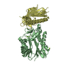
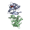
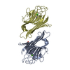
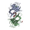
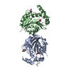
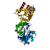
- PDB-5h65: Crystal structure of human POT1 and TPP1 -
+
Open data
ID or keywords:
Loading...
-
Basic information
Entry
Database: PDB / ID: 5h65
Title
Crystal structure of human POT1 and TPP1
Components
Adrenocortical dysplasia protein homolog
Protection of telomeres protein 1
Keywords
DNA BINDING PROTEIN / telomere / OB fold
Function / homology
Function and homology information
positive regulation of DNA strand elongation / positive regulation of telomeric D-loop disassembly / G-rich single-stranded DNA binding / telomere assembly / segmentation / regulation of double-strand break repair via nonhomologous end joining / urogenital system development / 8-hydroxy-2'-deoxyguanosine DNA binding / telomeric D-loop binding / DEAD/H-box RNA helicase binding ...positive regulation of DNA strand elongation / positive regulation of telomeric D-loop disassembly / G-rich single-stranded DNA binding / telomere assembly / segmentation / regulation of double-strand break repair via nonhomologous end joining / urogenital system development / 8-hydroxy-2'-deoxyguanosine DNA binding / telomeric D-loop binding / DEAD/H-box RNA helicase binding / protection from non-homologous end joining at telomere / establishment of protein localization to telomere / regulation of establishment of protein localization to telomere / telomerase inhibitor activity / regulation of telomere maintenance via telomerase / telomeric D-loop disassembly / shelterin complex / Telomere C-strand synthesis initiation / single-stranded telomeric DNA binding / Telomere C-strand (Lagging Strand) Synthesis / G-rich strand telomeric DNA binding / nuclear telomere cap complex / telomere capping / Polymerase switching on the C-strand of the telomere / telomerase holoenzyme complex / embryonic limb morphogenesis / Processive synthesis on the C-strand of the telomere / Removal of the Flap Intermediate from the C-strand / protein localization to chromosome, telomeric region / telomeric repeat DNA binding / negative regulation of telomere maintenance via telomerase / positive regulation of telomere maintenance / Telomere Extension By Telomerase / telomere maintenance via telomerase / positive regulation of telomere maintenance via telomerase / DNA polymerase binding / Packaging Of Telomere Ends / Recognition and association of DNA glycosylase with site containing an affected purine / Cleavage of the damaged purine / Recognition and association of DNA glycosylase with site containing an affected pyrimidine / Cleavage of the damaged pyrimidine / telomere maintenance / Inhibition of DNA recombination at telomere / Meiotic synapsis / skeletal system development / intracellular protein transport / DNA Damage/Telomere Stress Induced Senescence / chromosome, telomeric region / nuclear body / protein-containing complex binding / nucleoplasm Similarity search - Function
: / Protection of telomeres protein 1 (POT1), C-terminal insertion domain / Protection of telomeres protein 1, ssDNA-binding domain / ssDNA-binding domain of telomere protection protein / Protection of telomeres protein 1 / Adrenocortical dysplasia protein / Telomeric single stranded DNA binding POT1/Cdc13 / Telomeric single stranded DNA binding POT1/CDC13 / Telomeric single stranded DNA binding POT1/CDC13 / Shelterin complex subunit TPP1/Est3 ...: / Protection of telomeres protein 1 (POT1), C-terminal insertion domain / Protection of telomeres protein 1, ssDNA-binding domain / ssDNA-binding domain of telomere protection protein / Protection of telomeres protein 1 / Adrenocortical dysplasia protein / Telomeric single stranded DNA binding POT1/Cdc13 / Telomeric single stranded DNA binding POT1/CDC13 / Telomeric single stranded DNA binding POT1/CDC13 / Shelterin complex subunit TPP1/Est3 / Shelterin complex subunit, TPP1/ACD / Nucleic acid-binding, OB-fold Similarity search - Domain/homology
In the structure databanks used in Yorodumi, some data are registered as the other names, "COVID-19 virus" and "2019-nCoV". Here are the details of the virus and the list of structure data.
Jan 31, 2019. EMDB accession codes are about to change! (news from PDBe EMDB page)
EMDB accession codes are about to change! (news from PDBe EMDB page)
The allocation of 4 digits for EMDB accession codes will soon come to an end. Whilst these codes will remain in use, new EMDB accession codes will include an additional digit and will expand incrementally as the available range of codes is exhausted. The current 4-digit format prefixed with “EMD-” (i.e. EMD-XXXX) will advance to a 5-digit format (i.e. EMD-XXXXX), and so on. It is currently estimated that the 4-digit codes will be depleted around Spring 2019, at which point the 5-digit format will come into force.
The EM Navigator/Yorodumi systems omit the EMD- prefix.
Related info.:Q: What is EMD? / ID/Accession-code notation in Yorodumi/EM Navigator
Yorodumi is a browser for structure data from EMDB, PDB, SASBDB, etc.
This page is also the successor to EM Navigator detail page, and also detail information page/front-end page for Omokage search.
The word "yorodu" (or yorozu) is an old Japanese word meaning "ten thousand". "mi" (miru) is to see.
Related info.:EMDB / PDB / SASBDB / Comparison of 3 databanks / Yorodumi Search / Aug 31, 2016. New EM Navigator & Yorodumi / Yorodumi Papers / Jmol/JSmol / Function and homology information / Changes in new EM Navigator and Yorodumi
 Movie
Movie Controller


 Open data
 Basic information
 Components
 Keywords
 Function and homology information
 Homo sapiens (human)
 X-RAY DIFFRACTION /
  Authors
 Citation
 Structure visualization
 Downloads & links
 Other downloads
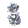


 PDBj
PDBj


 Assembly
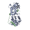


 Mass: 65.409 Da / Num. of mol.: 1 / Source method: obtained synthetically / Formula: Zn
 Mass: 18.015 Da / Num. of mol.: 140 / Source method: isolated from a natural source / Formula: H2O
 Sample preparation
 / Beamline: BL18U1 / Wavelength: 0.97876 Å
 Processing