 Movie
Movie Controller


+ Open data

- Basic information
| Entry | Database: PDB / ID: 3vcf | ||||||
|---|---|---|---|---|---|---|---|
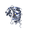
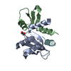
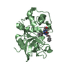
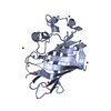
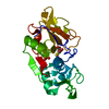
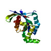
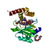
| Title | SSV1 integrase C-terminal catalytic domain (174-335aa) | ||||||
 Components Components | Probable integrase | ||||||
 Keywords Keywords | RECOMBINATION / catalyzes site-specific integration | ||||||
| Function / homology |  Function and homology information Function and homology informationDNA integration / viral genome integration into host DNA / establishment of integrated proviral latency / Transferases; Transferring phosphorus-containing groups; Nucleotidyltransferases / transferase activity / DNA recombination / Hydrolases; Acting on ester bonds / hydrolase activity / symbiont entry into host cell / DNA binding Similarity search - Function | ||||||
| Biological species |   Sulfolobus virus 1 Sulfolobus virus 1 | ||||||
| Method |  X-RAY DIFFRACTION / SYNCHROTRON / MOLECULAR REPLACEMENT / Resolution: 2.7 Å X-RAY DIFFRACTION / SYNCHROTRON / MOLECULAR REPLACEMENT / Resolution: 2.7 Å | ||||||
 Authors Authors | Ouyang, S. / Liang, W. / Huang, L. / Liu, Z.-J. | ||||||
 Citation Citation | Journal: Acta Crystallogr.,Sect.D / Year: 2012 Title: Structural and functional characterization of the C-terminal catalytic domain of SSV1 integrase. Authors: Zhan, Z. / Ouyang, S. / Liang, W. / Zhang, Z. / Liu, Z.J. / Huang, L. | ||||||
| History |
|
- Structure visualization
| Structure viewer | Molecule: MolmilJmol/JSmol |
|---|
- Downloads & links
-Download
| PDBx/mmCIF format | 3vcf.cif.gz | 77.5 KB | Display | PDBx/mmCIF format |
|---|---|---|---|---|
| PDB format | pdb3vcf.ent.gz | 58.3 KB | Display | PDB format |
| PDBx/mmJSON format | 3vcf.json.gz | Tree view | PDBx/mmJSON format | |
| Others |  Other downloads Other downloads |
-Validation report
| Arichive directory | https://data.pdbj.org/pub/pdb/validation_reports/vc/3vcfftp://data.pdbj.org/pub/pdb/validation_reports/vc/3vcf | HTTPS FTP |
|---|
-Related structure data


-Links
 PDBj
PDBj


- Assembly
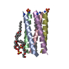
| Deposited unit | 
| ||||||||||||
|---|---|---|---|---|---|---|---|---|---|---|---|---|---|
| 1 | 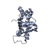
| ||||||||||||
| 2 | 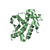
| ||||||||||||
| Unit cell |
| ||||||||||||
| Components on special symmetry positions |
| ||||||||||||
| Noncrystallographic symmetry (NCS) | NCS oper:
|
-Components
| #1: Protein | Mass: 19253.510 Da / Num. of mol.: 2 / Fragment: C-terminal domain (residues 174-335) Source method: isolated from a genetically manipulated source Source: (gene. exp.) Sulfolobus virus 1 / Gene: d335 / Plasmid: pET30a(+) / Production host:  #2: Water | ChemComp-HOH / |  Mass: 18.015 Da / Num. of mol.: 120 / Source method: isolated from a natural source / Formula: H2O Mass: 18.015 Da / Num. of mol.: 120 / Source method: isolated from a natural source / Formula: H2OHas protein modification | Y | |
|---|
-Experimental details
-Experiment
| Experiment | Method: X-RAY DIFFRACTION / Number of used crystals: 1 |
|---|
- Sample preparation
| Crystal | Density Matthews: 3.8 Å3/Da / Density % sol: 67.63 % |
|---|---|
| Crystal grow | Temperature: 289 K / Method: vapor diffusion, hanging drop / pH: 8.2 Details: 0.056M NaH2PO4, 1.35M K2HPO4, pH 8.2, VAPOR DIFFUSION, HANGING DROP, temperature 289K |
-Data collection
| Diffraction | Mean temperature: 100 K |
|---|---|
| Diffraction source | Source: SYNCHROTRON / Site: Photon Factory  / Beamline: BL-17A / Wavelength: 1 Å / Beamline: BL-17A / Wavelength: 1 Å |
| Detector | Type: ADSC QUANTUM 315 / Detector: CCD / Date: Feb 22, 2011 |
| Radiation | Monochromator: GRAPHITE / Protocol: SINGLE WAVELENGTH / Monochromatic (M) / Laue (L): M / Scattering type: x-ray |
| Radiation wavelength | Wavelength: 1 Å / Relative weight: 1 |
| Reflection | Resolution: 2.7→34.11 Å / Num. obs: 16941 / % possible obs: 99.9 % / Observed criterion σ(F): 2 / Observed criterion σ(I): 2 / Biso Wilson estimate: 31.45 Å2 |
| Reflection shell | Resolution: 2.7→2.95 Å / % possible all: 99.9 |
- Processing
| Software |
| ||||||||||||||||||||||||||||||||||||||||||
|---|---|---|---|---|---|---|---|---|---|---|---|---|---|---|---|---|---|---|---|---|---|---|---|---|---|---|---|---|---|---|---|---|---|---|---|---|---|---|---|---|---|---|---|
| Refinement | Method to determine structure: MOLECULAR REPLACEMENT / Resolution: 2.7→34.11 Å / Occupancy max: 1 / Occupancy min: 0.44 / FOM work R set: 0.8515 / SU ML: 0.28 / Cross valid method: THROUGHOUT / σ(F): 1.34 / Phase error: 20.99 / Stereochemistry target values: ML
| ||||||||||||||||||||||||||||||||||||||||||
| Solvent computation | Shrinkage radii: 0.98 Å / VDW probe radii: 1.2 Å / Solvent model: FLAT BULK SOLVENT MODEL / Bsol: 26.172 Å2 / ksol: 0.362 e/Å3 | ||||||||||||||||||||||||||||||||||||||||||
| Displacement parameters | Biso max: 93.56 Å2 / Biso mean: 28.1218 Å2 / Biso min: 7.38 Å2
| ||||||||||||||||||||||||||||||||||||||||||
| Refinement step | Cycle: LAST / Resolution: 2.7→34.11 Å
| ||||||||||||||||||||||||||||||||||||||||||
| Refine LS restraints |
| ||||||||||||||||||||||||||||||||||||||||||
| LS refinement shell | Refine-ID: X-RAY DIFFRACTION / Total num. of bins used: 6 / % reflection obs: 100 %
|
