 Movie
Movie Controller

[English] 日本語
 Yorodumi
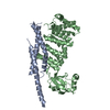
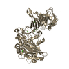
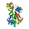
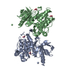
- PDB-2uv2: Crystal Structure Of Human Ste20-Like Kinase Bound To 4-(4-(5- Cy... -
+ Open data

- Basic information
| Entry | Database: PDB / ID: 2uv2 | |||||||||
|---|---|---|---|---|---|---|---|---|---|---|
| Title | Crystal Structure Of Human Ste20-Like Kinase Bound To 4-(4-(5- Cyclopropyl-1H-pyrazol-3-ylamino)-quinazolin-2-ylamino)-phenyl)- acetonitrile | |||||||||
 Components Components | STE20-LIKE SERINE-THREONINE KINASE | |||||||||
 Keywords Keywords | TRANSFERASE / SERINE/THREONINE-PROTEIN KINASE / ATP-BINDING / PHOSPHORYLATION / MUSCLE DEVELOPMENT / KINASE / APOPTOSIS / GERMINAL CENTRE KINASE / SERINE- THREONINE KINASE 2 / NUCLEOTIDE-BINDING / SERINE-THREONINE-PROTEIN KINASE | |||||||||
| Function / homology |  Function and homology information Function and homology informationregulation of focal adhesion assembly / RHOB GTPase cycle / RHOC GTPase cycle / cell leading edge / RHOA GTPase cycle / cytoplasmic microtubule organization / regulation of cell migration / protein autophosphorylation / regulation of apoptotic process / non-specific serine/threonine protein kinase ...regulation of focal adhesion assembly / RHOB GTPase cycle / RHOC GTPase cycle / cell leading edge / RHOA GTPase cycle / cytoplasmic microtubule organization / regulation of cell migration / protein autophosphorylation / regulation of apoptotic process / non-specific serine/threonine protein kinase / intracellular signal transduction / cadherin binding / protein serine kinase activity / protein serine/threonine kinase activity / apoptotic process / perinuclear region of cytoplasm / protein homodimerization activity / extracellular exosome / ATP binding / identical protein binding / cytoplasm / cytosol Similarity search - Function | |||||||||
| Biological species |  HOMO SAPIENS (human) HOMO SAPIENS (human) | |||||||||
| Method |  X-RAY DIFFRACTION / SYNCHROTRON / MOLECULAR REPLACEMENT / Resolution: 2.3 Å X-RAY DIFFRACTION / SYNCHROTRON / MOLECULAR REPLACEMENT / Resolution: 2.3 Å | |||||||||
 Authors Authors | Pike, A.C.W. / Rellos, P. / Fedorov, O. / Keates, T. / Salah, E. / Savitsky, P. / Papagrigoriou, E. / Bunkoczi, G. / Debreczeni, J.E. / von Delft, F. ...Pike, A.C.W. / Rellos, P. / Fedorov, O. / Keates, T. / Salah, E. / Savitsky, P. / Papagrigoriou, E. / Bunkoczi, G. / Debreczeni, J.E. / von Delft, F. / Arrowsmith, C.H. / Edwards, A. / Weigelt, J. / Sundstrom, M. / Knapp, S. | |||||||||
 Citation Citation | Journal: Embo J. / Year: 2008 Title: Activation Segment Dimerization: A Mechanism for Kinase Autophosphorylation of Non-Consensus Sites. Authors: Pike, A.C.W. / Rellos, P. / Niesen, F.H. / Turnbull, A. / Oliver, A.W. / Parker, S.A. / Turk, B.E. / Pearl, L.H. / Knapp, S. | |||||||||
| History |
|
- Structure visualization
| Structure viewer | Molecule: MolmilJmol/JSmol |
|---|
- Downloads & links
-Download
| PDBx/mmCIF format | 2uv2.cif.gz | 78.4 KB | Display | PDBx/mmCIF format |
|---|---|---|---|---|
| PDB format | pdb2uv2.ent.gz | 57 KB | Display | PDB format |
| PDBx/mmJSON format | 2uv2.json.gz | Tree view | PDBx/mmJSON format | |
| Others |  Other downloads Other downloads |
-Validation report
| Arichive directory | https://data.pdbj.org/pub/pdb/validation_reports/uv/2uv2ftp://data.pdbj.org/pub/pdb/validation_reports/uv/2uv2 | HTTPS FTP |
|---|
-Related structure data
| Related structure data |  2j51SC  2j7tC 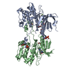 2j90C  2jflC 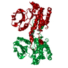 2jfmC S: Starting model for refinement C: citing same article ( |
|---|---|
| Similar structure data |


-Links
 PDBj
PDBj

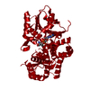
- Assembly
| Deposited unit | 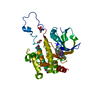
| ||||||||
|---|---|---|---|---|---|---|---|---|---|
| 1 | 
| ||||||||
| Unit cell |
| ||||||||
| Components on special symmetry positions |
|
-Components
| #1: Protein | Mass: 37162.070 Da / Num. of mol.: 1 / Fragment: KINASE DOMAIN, RESIDUES 19-320 / Mutation: YES Source method: isolated from a genetically manipulated source Source: (gene. exp.) HOMO SAPIENS (human) / Plasmid: PNIC28-BSA4 / Production host:  References: UniProt: Q9H2G2, non-specific serine/threonine protein kinase | ||||||||||
|---|---|---|---|---|---|---|---|---|---|---|---|
| #2: Chemical | ChemComp-EDO /   Mass: 62.068 Da / Num. of mol.: 5 / Source method: obtained synthetically / Formula: C2H6O2 Mass: 62.068 Da / Num. of mol.: 5 / Source method: obtained synthetically / Formula: C2H6O2#3: Chemical |   Mass: 58.082 Da / Num. of mol.: 2 / Source method: obtained synthetically / Formula: CNS Mass: 58.082 Da / Num. of mol.: 2 / Source method: obtained synthetically / Formula: CNS#4: Chemical | ChemComp-GVD / [ | 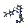  Mass: 381.433 Da / Num. of mol.: 1 / Source method: obtained synthetically / Formula: C22H19N7 Mass: 381.433 Da / Num. of mol.: 1 / Source method: obtained synthetically / Formula: C22H19N7#5: Water | ChemComp-HOH / |  Mass: 18.015 Da / Num. of mol.: 148 / Source method: isolated from a natural source / Formula: H2O Mass: 18.015 Da / Num. of mol.: 148 / Source method: isolated from a natural source / Formula: H2OCompound details | ENGINEERED | Sequence details | K25T MUTATION DUE TO CLONING ARTIFACT | |
-Experimental details
-Experiment
| Experiment | Method: X-RAY DIFFRACTION / Number of used crystals: 1 |
|---|
- Sample preparation
| Crystal | Density Matthews: 3.53 Å3/Da / Density % sol: 65 % |
|---|---|
| Crystal grow | pH: 6.5 Details: 18% PEG3350, 0.15M POTASSIUM THIOCYANATE, 10% ETHYLENE GLYCOL, 0.1M BISTRISPROPANE PH6.5, pH 6.50 |
-Data collection
| Diffraction | Mean temperature: 100 K |
|---|---|
| Diffraction source | Source: SYNCHROTRON / Site: SLS  / Beamline: X10SA / Wavelength: 0.9687 / Beamline: X10SA / Wavelength: 0.9687 |
| Detector | Type: MARRESEARCH / Detector: CCD / Date: Sep 1, 2006 |
| Radiation | Protocol: SINGLE WAVELENGTH / Monochromatic (M) / Laue (L): M / Scattering type: x-ray |
| Radiation wavelength | Wavelength: 0.9687 Å / Relative weight: 1 |
| Reflection | Resolution: 2.3→60 Å / Num. obs: 24390 / % possible obs: 99.3 % / Observed criterion σ(I): 0 / Redundancy: 8.4 % / Biso Wilson estimate: 50 Å2 / Rmerge(I) obs: 0.1 / Net I/σ(I): 14.2 |
| Reflection shell | Resolution: 2.3→2.42 Å / Redundancy: 6 % / Rmerge(I) obs: 0.86 / Mean I/σ(I) obs: 2.1 / % possible all: 95.2 |
- Processing
| Software |
| ||||||||||||||||||||||||||||||||||||||||||||||||||||||||||||||||||||||||||||||||||||||||||||||||||||||||||||||||||||||||||||||||||||||||||||||||||||||||||||||||||||||||||||||||||||||
|---|---|---|---|---|---|---|---|---|---|---|---|---|---|---|---|---|---|---|---|---|---|---|---|---|---|---|---|---|---|---|---|---|---|---|---|---|---|---|---|---|---|---|---|---|---|---|---|---|---|---|---|---|---|---|---|---|---|---|---|---|---|---|---|---|---|---|---|---|---|---|---|---|---|---|---|---|---|---|---|---|---|---|---|---|---|---|---|---|---|---|---|---|---|---|---|---|---|---|---|---|---|---|---|---|---|---|---|---|---|---|---|---|---|---|---|---|---|---|---|---|---|---|---|---|---|---|---|---|---|---|---|---|---|---|---|---|---|---|---|---|---|---|---|---|---|---|---|---|---|---|---|---|---|---|---|---|---|---|---|---|---|---|---|---|---|---|---|---|---|---|---|---|---|---|---|---|---|---|---|---|---|---|---|
| Refinement | Method to determine structure: MOLECULAR REPLACEMENT Starting model: PDB ENTRY 2J51 Resolution: 2.3→60 Å / Cor.coef. Fo:Fc: 0.952 / Cor.coef. Fo:Fc free: 0.931 / SU B: 10.504 / SU ML: 0.141 / TLS residual ADP flag: LIKELY RESIDUAL / Cross valid method: THROUGHOUT / ESU R: 0.199 / ESU R Free: 0.179 / Stereochemistry target values: MAXIMUM LIKELIHOOD / Details: HYDROGENS HAVE BEEN ADDED IN THE RIDING POSITIONS.
| ||||||||||||||||||||||||||||||||||||||||||||||||||||||||||||||||||||||||||||||||||||||||||||||||||||||||||||||||||||||||||||||||||||||||||||||||||||||||||||||||||||||||||||||||||||||
| Solvent computation | Ion probe radii: 0.8 Å / Shrinkage radii: 0.8 Å / VDW probe radii: 1.4 Å / Solvent model: MASK | ||||||||||||||||||||||||||||||||||||||||||||||||||||||||||||||||||||||||||||||||||||||||||||||||||||||||||||||||||||||||||||||||||||||||||||||||||||||||||||||||||||||||||||||||||||||
| Displacement parameters | Biso mean: 44.92 Å2
| ||||||||||||||||||||||||||||||||||||||||||||||||||||||||||||||||||||||||||||||||||||||||||||||||||||||||||||||||||||||||||||||||||||||||||||||||||||||||||||||||||||||||||||||||||||||
| Refinement step | Cycle: LAST / Resolution: 2.3→60 Å
| ||||||||||||||||||||||||||||||||||||||||||||||||||||||||||||||||||||||||||||||||||||||||||||||||||||||||||||||||||||||||||||||||||||||||||||||||||||||||||||||||||||||||||||||||||||||
| Refine LS restraints |
|