 Movie
Movie Controller


+ Open data

- Basic information
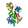
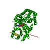
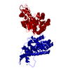
| Entry | Database: PDB / ID: 6xf2 | ||||||
|---|---|---|---|---|---|---|---|
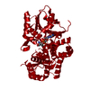
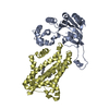
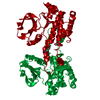
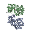
| Title | Nesprin-1G (aa2070-2200)-FHOD1(aa1-339) complex, H. sapiens | ||||||
 Components Components |
| ||||||
 Keywords Keywords | STRUCTURAL PROTEIN / spectrin repeat / FH3 domain / armadillo repeat / scaffold protein / nuclear positioning / transmembrane / actin-associated / nuclear line | ||||||
| Function / homology |  Function and homology information Function and homology informationnuclear matrix anchoring at nuclear membrane / cytoskeleton-nuclear membrane anchor activity / meiotic nuclear membrane microtubule tethering complex / regulation of cilium assembly / muscle cell differentiation / lamin binding / bleb / regulation of stress fiber assembly / nucleus organization / centrosome localization ...nuclear matrix anchoring at nuclear membrane / cytoskeleton-nuclear membrane anchor activity / meiotic nuclear membrane microtubule tethering complex / regulation of cilium assembly / muscle cell differentiation / lamin binding / bleb / regulation of stress fiber assembly / nucleus organization / centrosome localization / nuclear migration / Golgi organization / nuclear outer membrane / intercalated disc / positive regulation of stress fiber assembly / Meiotic synapsis / actin filament organization / sarcoplasmic reticulum / sarcomere / P-body / actin filament binding / nuclear envelope / actin binding / nuclear membrane / spermatogenesis / cytoskeleton / postsynaptic membrane / nucleolus / Golgi apparatus / enzyme binding / protein homodimerization activity / positive regulation of transcription by RNA polymerase II / RNA binding / nucleoplasm / membrane / identical protein binding / nucleus / cytosol / cytoplasm Similarity search - Function | ||||||
| Biological species |  Homo sapiens (human) Homo sapiens (human) | ||||||
| Method |  X-RAY DIFFRACTION / SYNCHROTRON / MOLECULAR REPLACEMENT / Resolution: 7.11 Å X-RAY DIFFRACTION / SYNCHROTRON / MOLECULAR REPLACEMENT / Resolution: 7.11 Å | ||||||
 Authors Authors | Lim, S.M. / Schwartz, T.U. | ||||||
| Funding support |  United States, 1items United States, 1items
| ||||||
 Citation Citation | Journal: Structure / Year: 2021 Title: Structures of FHOD1-Nesprin1/2 complexes reveal alternate binding modes for the FH3 domain of formins. Authors: Lim, S.M. / Cruz, V.E. / Antoku, S. / Gundersen, G.G. / Schwartz, T.U. | ||||||
| History |
|
- Structure visualization
| Structure viewer | Molecule: MolmilJmol/JSmol |
|---|
- Downloads & links
-Download
| PDBx/mmCIF format | 6xf2.cif.gz | 190.5 KB | Display | PDBx/mmCIF format |
|---|---|---|---|---|
| PDB format | pdb6xf2.ent.gz | 126.1 KB | Display | PDB format |
| PDBx/mmJSON format | 6xf2.json.gz | Tree view | PDBx/mmJSON format | |
| Others |  Other downloads Other downloads |
-Validation report
| Arichive directory | https://data.pdbj.org/pub/pdb/validation_reports/xf/6xf2ftp://data.pdbj.org/pub/pdb/validation_reports/xf/6xf2 | HTTPS FTP |
|---|
-Related structure data
| Related structure data | 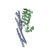 6xf1SC S: Starting model for refinement C: citing same article ( |
|---|---|
| Similar structure data |


-Links
 PDBj
PDBj
- Assembly
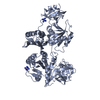
| Deposited unit | 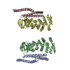
| ||||||||||||||||||||||||||||||||||||||||||||||||||||||||||||||||||||||||
|---|---|---|---|---|---|---|---|---|---|---|---|---|---|---|---|---|---|---|---|---|---|---|---|---|---|---|---|---|---|---|---|---|---|---|---|---|---|---|---|---|---|---|---|---|---|---|---|---|---|---|---|---|---|---|---|---|---|---|---|---|---|---|---|---|---|---|---|---|---|---|---|---|---|
| 1 | 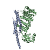
| ||||||||||||||||||||||||||||||||||||||||||||||||||||||||||||||||||||||||
| 2 | 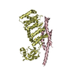
| ||||||||||||||||||||||||||||||||||||||||||||||||||||||||||||||||||||||||
| Unit cell |
| ||||||||||||||||||||||||||||||||||||||||||||||||||||||||||||||||||||||||
| Noncrystallographic symmetry (NCS) | NCS domain:
NCS domain segments:
NCS ensembles :
NCS oper:
|
-Components
| #1: Protein | Mass: 15259.411 Da / Num. of mol.: 2 Source method: isolated from a genetically manipulated source Source: (gene. exp.) Homo sapiens (human) / Gene: SYNE1, C6orf98, KIAA0796, KIAA1262, KIAA1756, MYNE1 / Production host:  #2: Protein | Mass: 35489.484 Da / Num. of mol.: 2 Source method: isolated from a genetically manipulated source Source: (gene. exp.) Homo sapiens (human) / Gene: FHOD1, FHOS, FHOS1 / Production host: |
|---|
-Experimental details
-Experiment
| Experiment | Method: X-RAY DIFFRACTION / Number of used crystals: 1 |
|---|
- Sample preparation
| Crystal | Density Matthews: 3.75 Å3/Da / Density % sol: 65.27 % |
|---|---|
| Crystal grow | Temperature: 292 K / Method: vapor diffusion, sitting drop Details: 0.1 M HEPES/NaOH pH 7.0, 0.2 M sodium thiocyanate, 40% 5/4 pentaerythritol propoxylate |
-Data collection
| Diffraction | Mean temperature: 80 K / Serial crystal experiment: N |
|---|---|
| Diffraction source | Source: SYNCHROTRON / Site: APS / Beamline: 24-ID-C / Wavelength: 0.987 Å |
| Detector | Type: ADSC QUANTUM 315 / Detector: CCD / Date: Nov 1, 2018 |
| Radiation | Protocol: SINGLE WAVELENGTH / Monochromatic (M) / Laue (L): M / Scattering type: x-ray |
| Radiation wavelength | Wavelength: 0.987 Å / Relative weight: 1 |
| Reflection | Resolution: 7.1→60.58 Å / Num. obs: 2378 / % possible obs: 98.2 % / Redundancy: 6 % / Biso Wilson estimate: 464.08 Å2 / CC1/2: 0.98 / Rpim(I) all: 0.063 / Net I/σ(I): 15.6 |
| Reflection shell | Resolution: 7.1→7.35 Å / Num. unique obs: 232 / CC1/2: 0.63 |
- Processing
| Software |
| ||||||||||||||||||||||||
|---|---|---|---|---|---|---|---|---|---|---|---|---|---|---|---|---|---|---|---|---|---|---|---|---|---|
| Refinement | Method to determine structure: MOLECULAR REPLACEMENT Starting model: 6XF1 Resolution: 7.11→60.58 Å / SU ML: 1.2952 / Cross valid method: FREE R-VALUE / σ(F): 1.35 / Phase error: 37.3238 Stereochemistry target values: GeoStd + Monomer Library + CDL v1.2
| ||||||||||||||||||||||||
| Solvent computation | Shrinkage radii: 0.9 Å / VDW probe radii: 1.11 Å / Solvent model: FLAT BULK SOLVENT MODEL | ||||||||||||||||||||||||
| Displacement parameters | Biso mean: 320.7 Å2 | ||||||||||||||||||||||||
| Refinement step | Cycle: LAST / Resolution: 7.11→60.58 Å
| ||||||||||||||||||||||||
| Refine LS restraints |
| ||||||||||||||||||||||||
| Refine LS restraints NCS |
| ||||||||||||||||||||||||
| LS refinement shell |
|
