 Movie
Movie Controller

[English] 日本語
 Yorodumi
- PDB-4oh7: Crystal structure of Ornithine carbamoyltransferase from Brucella... -
+ Open data

- Basic information
| Entry | Database: PDB / ID: 4oh7 | ||||||
|---|---|---|---|---|---|---|---|
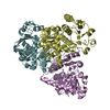
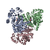
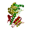
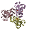
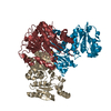
| Title | Crystal structure of Ornithine carbamoyltransferase from Brucella melitensis | ||||||
 Components Components | Ornithine carbamoyltransferase | ||||||
 Keywords Keywords | TRANSFERASE / SSGCID / structural genomics / Seattle Structural Genomics Center for Infectious Disease | ||||||
| Function / homology |  Function and homology information Function and homology informationornithine carbamoyltransferase / ornithine carbamoyltransferase activity / L-citrulline biosynthetic process / : / amino acid binding / cytoplasm Similarity search - Function | ||||||
| Biological species |  Brucella melitensis (bacteria) Brucella melitensis (bacteria) | ||||||
| Method |  X-RAY DIFFRACTION / SYNCHROTRON / MOLECULAR REPLACEMENT / molecular replacement / Resolution: 1.5 Å X-RAY DIFFRACTION / SYNCHROTRON / MOLECULAR REPLACEMENT / molecular replacement / Resolution: 1.5 Å | ||||||
 Authors Authors | Seattle Structural Genomics Center for Infectious Disease (SSGCID) / Seattle Structural Genomics Center for Infectious Disease (SSGCID) | ||||||
 Citation Citation | Journal: To be Published Title: Crystal structure of Ornithine carbamoyltransferase from Brucella melitensis Authors: Abendroth, J. / Lorimer, D. / Edwards, T.E. | ||||||
| History |
|
- Structure visualization
| Structure viewer | Molecule: MolmilJmol/JSmol |
|---|
- Downloads & links
-Download
| PDBx/mmCIF format | 4oh7.cif.gz | 257.1 KB | Display | PDBx/mmCIF format |
|---|---|---|---|---|
| PDB format | pdb4oh7.ent.gz | 207.4 KB | Display | PDB format |
| PDBx/mmJSON format | 4oh7.json.gz | Tree view | PDBx/mmJSON format | |
| Others |  Other downloads Other downloads |
-Validation report
| Arichive directory | https://data.pdbj.org/pub/pdb/validation_reports/oh/4oh7ftp://data.pdbj.org/pub/pdb/validation_reports/oh/4oh7 | HTTPS FTP |
|---|
-Related structure data
| Similar structure data | |
|---|---|
| Other databases |
-Links
 PDBj
PDBj
- Assembly
| Deposited unit | 
| |||||||||
|---|---|---|---|---|---|---|---|---|---|---|
| 1 | 
| |||||||||
| 2 | 
| |||||||||
| Unit cell |
| |||||||||
| Components on special symmetry positions |
|
-Components
| #1: Protein | Mass: 35313.090 Da / Num. of mol.: 2 Source method: isolated from a genetically manipulated source Source: (gene. exp.) Brucella melitensis (bacteria) / Strain: biotype 2 (strain ATCC 23457) / Gene: arcB, argF, BMEA_A0337 / Plasmid: BrmeB.00183.a.B1 / Production host: #2: Chemical | ChemComp-EDO /   Mass: 62.068 Da / Num. of mol.: 4 / Source method: obtained synthetically / Formula: C2H6O2 Mass: 62.068 Da / Num. of mol.: 4 / Source method: obtained synthetically / Formula: C2H6O2#3: Chemical |   Mass: 35.453 Da / Num. of mol.: 2 / Source method: obtained synthetically / Formula: Cl Mass: 35.453 Da / Num. of mol.: 2 / Source method: obtained synthetically / Formula: Cl#4: Water | ChemComp-HOH / |  Mass: 18.015 Da / Num. of mol.: 711 / Source method: isolated from a natural source / Formula: H2O Mass: 18.015 Da / Num. of mol.: 711 / Source method: isolated from a natural source / Formula: H2O |
|---|
-Experimental details
-Experiment
| Experiment | Method: X-RAY DIFFRACTION / Number of used crystals: 1 |
|---|
- Sample preparation
| Crystal | Density Matthews: 2.28 Å3/Da / Density % sol: 46.08 % |
|---|---|
| Crystal grow | Temperature: 290 K / Method: vapor diffusion, sitting drop / pH: 5.5 Details: BrmeB.00183.a.B1.PS01877 at 20.3 mg/mL, RigakuReagents JCSG+ screen, h8: 25% PEG3350, 200 mM sodium chloride, 100 mM Bis-Tris-HCl, pH 5.5, cryoprotection: 15% ethylene glycol in two steps, ...Details: BrmeB.00183.a.B1.PS01877 at 20.3 mg/mL, RigakuReagents JCSG+ screen, h8: 25% PEG3350, 200 mM sodium chloride, 100 mM Bis-Tris-HCl, pH 5.5, cryoprotection: 15% ethylene glycol in two steps, VAPOR DIFFUSION, SITTING DROP, temperature 290K |
-Data collection
| Diffraction | Mean temperature: 100 K | |||||||||||||||||||||||||||||||||||||||||||||||||||||||||||||||||||||||||||||||||||||||||||||||||||||||||||||||||||||||||||||||||||||||||||||||||||
|---|---|---|---|---|---|---|---|---|---|---|---|---|---|---|---|---|---|---|---|---|---|---|---|---|---|---|---|---|---|---|---|---|---|---|---|---|---|---|---|---|---|---|---|---|---|---|---|---|---|---|---|---|---|---|---|---|---|---|---|---|---|---|---|---|---|---|---|---|---|---|---|---|---|---|---|---|---|---|---|---|---|---|---|---|---|---|---|---|---|---|---|---|---|---|---|---|---|---|---|---|---|---|---|---|---|---|---|---|---|---|---|---|---|---|---|---|---|---|---|---|---|---|---|---|---|---|---|---|---|---|---|---|---|---|---|---|---|---|---|---|---|---|---|---|---|---|---|---|
| Diffraction source | Source: SYNCHROTRON / Site: ALS  / Beamline: 5.0.2 / Wavelength: 1 Å / Beamline: 5.0.2 / Wavelength: 1 Å | |||||||||||||||||||||||||||||||||||||||||||||||||||||||||||||||||||||||||||||||||||||||||||||||||||||||||||||||||||||||||||||||||||||||||||||||||||
| Detector | Type: ADSC QUANTUM 315r / Detector: CCD / Date: Dec 22, 2013 | |||||||||||||||||||||||||||||||||||||||||||||||||||||||||||||||||||||||||||||||||||||||||||||||||||||||||||||||||||||||||||||||||||||||||||||||||||
| Radiation | Monochromator: double crystal Si(111) / Protocol: SINGLE WAVELENGTH / Monochromatic (M) / Laue (L): M / Scattering type: x-ray | |||||||||||||||||||||||||||||||||||||||||||||||||||||||||||||||||||||||||||||||||||||||||||||||||||||||||||||||||||||||||||||||||||||||||||||||||||
| Radiation wavelength | Wavelength: 1 Å / Relative weight: 1 | |||||||||||||||||||||||||||||||||||||||||||||||||||||||||||||||||||||||||||||||||||||||||||||||||||||||||||||||||||||||||||||||||||||||||||||||||||
| Reflection | Resolution: 1.5→50 Å / Num. all: 100819 / Num. obs: 100352 / % possible obs: 99.5 % / Observed criterion σ(F): 0 / Observed criterion σ(I): -3 / Redundancy: 3.6 % / Biso Wilson estimate: 23.829 Å2 / Rmerge(I) obs: 0.044 / Net I/σ(I): 16.38 | |||||||||||||||||||||||||||||||||||||||||||||||||||||||||||||||||||||||||||||||||||||||||||||||||||||||||||||||||||||||||||||||||||||||||||||||||||
| Reflection shell |
|
-Phasing
| Phasing | Method: molecular replacement |
|---|---|
| Phasing MR | Model details: Phaser MODE: MR_AUTO |
- Processing
| Software |
| |||||||||||||||||||||||||||||||||||||||||||||||||||||||||||||||||||||||||||
|---|---|---|---|---|---|---|---|---|---|---|---|---|---|---|---|---|---|---|---|---|---|---|---|---|---|---|---|---|---|---|---|---|---|---|---|---|---|---|---|---|---|---|---|---|---|---|---|---|---|---|---|---|---|---|---|---|---|---|---|---|---|---|---|---|---|---|---|---|---|---|---|---|---|---|---|---|
| Refinement | Method to determine structure: MOLECULAR REPLACEMENT Starting model: MODEL IN DIFFERENT SPACE GROUP OBTAINED FROM IODIDE PHASING Resolution: 1.5→50 Å / Cor.coef. Fo:Fc: 0.971 / Cor.coef. Fo:Fc free: 0.96 / WRfactor Rfree: 0.1797 / WRfactor Rwork: 0.1519 / FOM work R set: 0.8895 / SU B: 2.42 / SU ML: 0.045 / SU R Cruickshank DPI: 0.0642 / SU Rfree: 0.0666 / Isotropic thermal model: ISOTROPIC, TLS / Cross valid method: THROUGHOUT / σ(F): 0 / ESU R: 0.064 / ESU R Free: 0.067 / Stereochemistry target values: MAXIMUM LIKELIHOOD / Details: HYDROGENS HAVE BEEN ADDED IN THE RIDING POSITIONS
| |||||||||||||||||||||||||||||||||||||||||||||||||||||||||||||||||||||||||||
| Solvent computation | Ion probe radii: 0.8 Å / Shrinkage radii: 0.8 Å / VDW probe radii: 1.2 Å / Solvent model: MASK | |||||||||||||||||||||||||||||||||||||||||||||||||||||||||||||||||||||||||||
| Displacement parameters | Biso max: 59.76 Å2 / Biso mean: 19.044 Å2 / Biso min: 7.87 Å2
| |||||||||||||||||||||||||||||||||||||||||||||||||||||||||||||||||||||||||||
| Refinement step | Cycle: LAST / Resolution: 1.5→50 Å
| |||||||||||||||||||||||||||||||||||||||||||||||||||||||||||||||||||||||||||
| Refine LS restraints |
| |||||||||||||||||||||||||||||||||||||||||||||||||||||||||||||||||||||||||||
| LS refinement shell | Resolution: 1.5→1.539 Å / Total num. of bins used: 20
| |||||||||||||||||||||||||||||||||||||||||||||||||||||||||||||||||||||||||||
| Refinement TLS params. | Method: refined / Refine-ID: X-RAY DIFFRACTION
| |||||||||||||||||||||||||||||||||||||||||||||||||||||||||||||||||||||||||||
| Refinement TLS group |
|
