 Movie
Movie Controller

[English] 日本語
 Yorodumi
- PDB-3e2p: Catalytic subunit of M. Jannaschii aspartate transcarbamoylase in... -
+ Open data

- Basic information
| Entry | Database: PDB / ID: 3e2p | ||||||
|---|---|---|---|---|---|---|---|
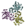
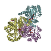
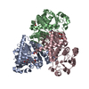
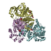
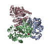
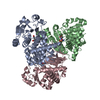
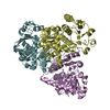
| Title | Catalytic subunit of M. Jannaschii aspartate transcarbamoylase in an orthorhombic crystal form | ||||||
 Components Components | Aspartate carbamoyltransferase | ||||||
 Keywords Keywords | TRANSFERASE / Aspartate Transcarbamoylase / ATCase / pyrimidine biosynthesis / thermostability / Methanococcus jannaschii | ||||||
| Function / homology |  Function and homology information Function and homology informationaspartate carbamoyltransferase / aspartate carbamoyltransferase activity / L-glutamine metabolic process / amino acid metabolic process / amino acid binding / 'de novo' UMP biosynthetic process / 'de novo' pyrimidine nucleobase biosynthetic process / cytoplasm Similarity search - Function | ||||||
| Biological species |   Methanocaldococcus jannaschii (archaea) Methanocaldococcus jannaschii (archaea) | ||||||
| Method |  X-RAY DIFFRACTION / SYNCHROTRON / MOLECULAR REPLACEMENT / Resolution: 3 Å X-RAY DIFFRACTION / SYNCHROTRON / MOLECULAR REPLACEMENT / Resolution: 3 Å | ||||||
 Authors Authors | Vitali, J. / Colaneri, M.J. | ||||||
 Citation Citation | Journal: Acta Crystallogr.,Sect.F / Year: 2008 Title: Structure of the catalytic trimer of Methanococcus jannaschii aspartate transcarbamoylase in an orthorhombic crystal form. Authors: Vitali, J. / Colaneri, M.J. #1: Journal: PROTEINS / Year: 2008Title: Crystal structure of the catalytic trimer of Methanococcus jannaschii aspartate transcarbamoylas Authors: Vitali, J. / Colaneri, M.J. / Kantrowitz, E. | ||||||
| History |
|
- Structure visualization
| Structure viewer | Molecule: MolmilJmol/JSmol |
|---|
- Downloads & links
-Download
| PDBx/mmCIF format | 3e2p.cif.gz | 652.7 KB | Display | PDBx/mmCIF format |
|---|---|---|---|---|
| PDB format | pdb3e2p.ent.gz | 558.6 KB | Display | PDB format |
| PDBx/mmJSON format | 3e2p.json.gz | Tree view | PDBx/mmJSON format | |
| Others |  Other downloads Other downloads |
-Validation report
| Arichive directory | https://data.pdbj.org/pub/pdb/validation_reports/e2/3e2pftp://data.pdbj.org/pub/pdb/validation_reports/e2/3e2p | HTTPS FTP |
|---|
-Related structure data
| Related structure data |  2rgwS S: Starting model for refinement |
|---|---|
| Similar structure data |


-Links
 PDBj
PDBj
- Assembly
| Deposited unit | 
| ||||||||
|---|---|---|---|---|---|---|---|---|---|
| 1 | 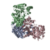
| ||||||||
| 2 | 
| ||||||||
| 3 | 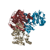
| ||||||||
| 4 | 
| ||||||||
| 5 | 
| ||||||||
| 6 | 
| ||||||||
| Unit cell |
|
-Components
| #1: Protein | Mass: 35210.621 Da / Num. of mol.: 12 / Fragment: catalytic subunit Source method: isolated from a genetically manipulated source Details: PSJS1240 was also co-transformed Source: (gene. exp.) Methanocaldococcus jannaschii (archaea)Gene: pyrB, MJ1581 / Plasmid: pEK406 / Production host:  #2: Chemical | ChemComp-SO4 /   Mass: 96.063 Da / Num. of mol.: 4 / Source method: obtained synthetically / Formula: SO4 Mass: 96.063 Da / Num. of mol.: 4 / Source method: obtained synthetically / Formula: SO4#3: Water | ChemComp-HOH / |  Mass: 18.015 Da / Num. of mol.: 500 / Source method: isolated from a natural source / Formula: H2O Mass: 18.015 Da / Num. of mol.: 500 / Source method: isolated from a natural source / Formula: H2O |
|---|
-Experimental details
-Experiment
| Experiment | Method: X-RAY DIFFRACTION / Number of used crystals: 2 |
|---|
- Sample preparation
| Crystal | Density Matthews: 2.77 Å3/Da / Density % sol: 55.52 % |
|---|---|
| Crystal grow | Temperature: 295 K / Method: vapor diffusion, sitting drop / pH: 8.5 Details: 2.5 M ammonium sulfate, 0.1 M Tris-HCl pH 8.5, VAPOR DIFFUSION, SITTING DROP, temperature 295K |
-Data collection
| Diffraction | Mean temperature: 100 K |
|---|---|
| Diffraction source | Source: SYNCHROTRON / Site: NSLS  / Beamline: X12B / Wavelength: 0.979 Å / Beamline: X12B / Wavelength: 0.979 Å |
| Detector | Type: ADSC QUANTUM 4 / Detector: CCD |
| Radiation | Monochromator: SI (111) crystal monochromator / Protocol: SINGLE WAVELENGTH / Monochromatic (M) / Laue (L): M / Scattering type: x-ray |
| Radiation wavelength | Wavelength: 0.979 Å / Relative weight: 1 |
| Reflection | Resolution: 3→40 Å / Num. obs: 92280 / % possible obs: 99.5 % / Redundancy: 4.4 % / Biso Wilson estimate: 73.3 Å2 / Rmerge(I) obs: 0.095 |
| Reflection shell | Resolution: 3→3.11 Å / Redundancy: 2.8 % / Rmerge(I) obs: 0.67 / Mean I/σ(I) obs: 1.4 / Num. unique all: 8844 / % possible all: 96.8 |
- Processing
| Software |
| |||||||||||||||||||||||||
|---|---|---|---|---|---|---|---|---|---|---|---|---|---|---|---|---|---|---|---|---|---|---|---|---|---|---|
| Refinement | Method to determine structure: MOLECULAR REPLACEMENT Starting model: 2RGW Resolution: 3→40 Å / Isotropic thermal model: isotropic / Cross valid method: THROUGHOUT / σ(F): 0 / Stereochemistry target values: Engh & Huber
| |||||||||||||||||||||||||
| Displacement parameters | Biso mean: 70 Å2 | |||||||||||||||||||||||||
| Refine analyze |
| |||||||||||||||||||||||||
| Refinement step | Cycle: LAST / Resolution: 3→40 Å
| |||||||||||||||||||||||||
| Refine LS restraints |
|
