 Movie
Movie Controller

[English] 日本語
 Yorodumi
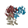
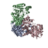
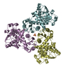
- PDB-4ekn: Structure of the catalytic chain of Methanococcus jannaschii Aspa... -
+ Open data

- Basic information
| Entry | Database: PDB / ID: 4ekn | ||||||
|---|---|---|---|---|---|---|---|
| Title | Structure of the catalytic chain of Methanococcus jannaschii Aspartate Transcarbamoylase in a hexagonal crystal form | ||||||
 Components Components | Aspartate carbamoyltransferase | ||||||
 Keywords Keywords | TRANSFERASE / ATCase / aspartate transcarbamoylase / pyrimidine biosynthesis / thermostability / substrate channeling | ||||||
| Function / homology |  Function and homology information Function and homology informationaspartate carbamoyltransferase / aspartate carbamoyltransferase activity / L-glutamine metabolic process / amino acid metabolic process / amino acid binding / 'de novo' UMP biosynthetic process / 'de novo' pyrimidine nucleobase biosynthetic process / cytoplasm Similarity search - Function | ||||||
| Biological species |   Methanocaldococcus jannaschii (archaea) Methanocaldococcus jannaschii (archaea) | ||||||
| Method |  X-RAY DIFFRACTION / SYNCHROTRON / MOLECULAR REPLACEMENT / Resolution: 2.4996 Å X-RAY DIFFRACTION / SYNCHROTRON / MOLECULAR REPLACEMENT / Resolution: 2.4996 Å | ||||||
 Authors Authors | Vitali, J. | ||||||
 Citation Citation | Journal: Acta Crystallogr.,Sect.F / Year: 2012 Title: Structure of the catalytic chain of Methanococcus jannaschii aspartate transcarbamoylase in a hexagonal crystal form: insights into the path of carbamoyl phosphate to the active site of the enzyme. Authors: Vitali, J. / Singh, A.K. / Soares, A.S. / Colaneri, M.J. | ||||||
| History |
|
- Structure visualization
| Structure viewer | Molecule: MolmilJmol/JSmol |
|---|
- Downloads & links
-Download
| PDBx/mmCIF format | 4ekn.cif.gz | 79.1 KB | Display | PDBx/mmCIF format |
|---|---|---|---|---|
| PDB format | pdb4ekn.ent.gz | 58.8 KB | Display | PDB format |
| PDBx/mmJSON format | 4ekn.json.gz | Tree view | PDBx/mmJSON format | |
| Others |  Other downloads Other downloads |
-Validation report
| Arichive directory | https://data.pdbj.org/pub/pdb/validation_reports/ek/4eknftp://data.pdbj.org/pub/pdb/validation_reports/ek/4ekn | HTTPS FTP |
|---|
-Related structure data
| Related structure data |  2rgwS S: Starting model for refinement |
|---|---|
| Similar structure data |


-Links
 PDBj
PDBj
- Assembly
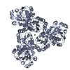
| Deposited unit | 
| |||||||||||||||||||||||||||
|---|---|---|---|---|---|---|---|---|---|---|---|---|---|---|---|---|---|---|---|---|---|---|---|---|---|---|---|---|
| 1 | 
| |||||||||||||||||||||||||||
| 2 | x 6
| |||||||||||||||||||||||||||
| Unit cell |
| |||||||||||||||||||||||||||
| Components on special symmetry positions |
| |||||||||||||||||||||||||||
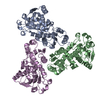
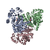
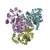
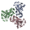
| Details | Trimer. A hexamer is also formed by pisa prediction but it is not known if it has biological relevance |
-Components
| #1: Protein | Mass: 35210.621 Da / Num. of mol.: 1 Source method: isolated from a genetically manipulated source Source: (gene. exp.) Methanocaldococcus jannaschii (archaea)Strain: ATCC 43067 / DSM 2661 / JAL-1 / JCM 10045 / NBRC 100440 Description: pSJS1240 was also cotransformed / Gene: MJ1581, pyrB / Plasmid: pEK406 / Production host:  | ||||||
|---|---|---|---|---|---|---|---|
| #2: Chemical | ChemComp-SO4 /   Mass: 96.063 Da / Num. of mol.: 4 / Source method: obtained synthetically / Formula: SO4 Mass: 96.063 Da / Num. of mol.: 4 / Source method: obtained synthetically / Formula: SO4#3: Chemical | ChemComp-K / |   Mass: 39.098 Da / Num. of mol.: 1 / Source method: obtained synthetically / Formula: K Mass: 39.098 Da / Num. of mol.: 1 / Source method: obtained synthetically / Formula: K#4: Chemical | ChemComp-GOL / |   Mass: 92.094 Da / Num. of mol.: 1 / Source method: obtained synthetically / Formula: C3H8O3 Mass: 92.094 Da / Num. of mol.: 1 / Source method: obtained synthetically / Formula: C3H8O3#5: Water | ChemComp-HOH / |  Mass: 18.015 Da / Num. of mol.: 145 / Source method: isolated from a natural source / Formula: H2O Mass: 18.015 Da / Num. of mol.: 145 / Source method: isolated from a natural source / Formula: H2O |
-Experimental details
-Experiment
| Experiment | Method: X-RAY DIFFRACTION / Number of used crystals: 1 |
|---|
- Sample preparation
| Crystal | Density Matthews: 2.63 Å3/Da / Density % sol: 53.21 % |
|---|---|
| Crystal grow | Temperature: 295 K / Method: vapor diffusion, sitting drop / pH: 7.5 Details: 2.0 M ammonium sulfate, 0.2 M potassium sodium tartrate tetrahydrate, and 0.1 M Tris-HCl pH 7.5. The protein was a mixture of catalytic and regulatory subunits at a molar ratio of 1:1 ...Details: 2.0 M ammonium sulfate, 0.2 M potassium sodium tartrate tetrahydrate, and 0.1 M Tris-HCl pH 7.5. The protein was a mixture of catalytic and regulatory subunits at a molar ratio of 1:1 concentrated to 11 mg/ml. Drops consisted of 2ul reservoir and 2.6 ul complex solution., VAPOR DIFFUSION, SITTING DROP, temperature 295K |
-Data collection
| Diffraction | Mean temperature: 100 K |
|---|---|
| Diffraction source | Source: SYNCHROTRON / Site: NSLS  / Beamline: X12C / Wavelength: 1.1 Å / Beamline: X12C / Wavelength: 1.1 Å |
| Detector | Type: ADSC QUANTUM 210 / Detector: CCD / Date: Mar 18, 2010 |
| Radiation | Monochromator: Si(111) crystal monochromator / Protocol: SINGLE WAVELENGTH / Monochromatic (M) / Laue (L): M / Scattering type: x-ray |
| Radiation wavelength | Wavelength: 1.1 Å / Relative weight: 1 |
| Reflection | Resolution: 2.4996→50 Å / Num. obs: 13471 / % possible obs: 98 % / Observed criterion σ(I): -3 / Redundancy: 3.3 % / Biso Wilson estimate: 32.4 Å2 / Rmerge(I) obs: 0.135 / Net I/σ(I): 9.3 |
| Reflection shell | Resolution: 2.4996→2.59 Å / Redundancy: 3.3 % / Rmerge(I) obs: 0.825 / Mean I/σ(I) obs: 1.62 / % possible all: 99.2 |
- Processing
| Software |
| ||||||||||||||||||||||||||||||||||||||||||||||||||||||||||||||||||||||||||||||||||||||||
|---|---|---|---|---|---|---|---|---|---|---|---|---|---|---|---|---|---|---|---|---|---|---|---|---|---|---|---|---|---|---|---|---|---|---|---|---|---|---|---|---|---|---|---|---|---|---|---|---|---|---|---|---|---|---|---|---|---|---|---|---|---|---|---|---|---|---|---|---|---|---|---|---|---|---|---|---|---|---|---|---|---|---|---|---|---|---|---|---|---|
| Refinement | Method to determine structure: MOLECULAR REPLACEMENT Starting model: PDB ENTRY of 2RGW chain D Resolution: 2.4996→41.985 Å / SU ML: 0.35 / Isotropic thermal model: isotropic / Cross valid method: THROUGHOUT / Phase error: 24.63 / Stereochemistry target values: ML
| ||||||||||||||||||||||||||||||||||||||||||||||||||||||||||||||||||||||||||||||||||||||||
| Solvent computation | Shrinkage radii: 0.83 Å / VDW probe radii: 1.1 Å / Solvent model: FLAT BULK SOLVENT MODEL / Bsol: 63.097 Å2 / ksol: 0.405 e/Å3 | ||||||||||||||||||||||||||||||||||||||||||||||||||||||||||||||||||||||||||||||||||||||||
| Displacement parameters | Biso mean: 38.5 Å2
| ||||||||||||||||||||||||||||||||||||||||||||||||||||||||||||||||||||||||||||||||||||||||
| Refinement step | Cycle: LAST / Resolution: 2.4996→41.985 Å
| ||||||||||||||||||||||||||||||||||||||||||||||||||||||||||||||||||||||||||||||||||||||||
| Refine LS restraints |
| ||||||||||||||||||||||||||||||||||||||||||||||||||||||||||||||||||||||||||||||||||||||||
| LS refinement shell | Refine-ID: X-RAY DIFFRACTION
|
