 Movie
Movie Controller

[English] 日本語
 Yorodumi
- PDB-6jl6: Crystal structure of aspartate transcarbamoylase from Trypanosoma... -
+ Open data

- Basic information
| Entry | Database: PDB / ID: 6jl6 | ||||||
|---|---|---|---|---|---|---|---|
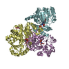
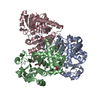
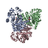
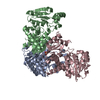
| Title | Crystal structure of aspartate transcarbamoylase from Trypanosoma cruzi in complex with phosphate (Pi). | ||||||
 Components Components | Aspartate carbamoyltransferase | ||||||
 Keywords Keywords | TRANSFERASE / ATCase / Pyrimidine metabolism | ||||||
| Function / homology |  Function and homology information Function and homology informationaspartate carbamoyltransferase / aspartate carbamoyltransferase activity / amino acid metabolic process / amino acid binding / 'de novo' UMP biosynthetic process / 'de novo' pyrimidine nucleobase biosynthetic process Similarity search - Function | ||||||
| Biological species |  | ||||||
| Method |  X-RAY DIFFRACTION / SYNCHROTRON / MOLECULAR REPLACEMENT / Resolution: 2 Å X-RAY DIFFRACTION / SYNCHROTRON / MOLECULAR REPLACEMENT / Resolution: 2 Å | ||||||
 Authors Authors | Matoba, K. / Shiba, T. / Nara, T. / Aoki, T. / Nagasaki, S. / Hayamizu, R. / Honma, T. / Tanaka, A. / Inoue, M. / Matsuoka, S. ...Matoba, K. / Shiba, T. / Nara, T. / Aoki, T. / Nagasaki, S. / Hayamizu, R. / Honma, T. / Tanaka, A. / Inoue, M. / Matsuoka, S. / Balogun, E.O. / Inaoka, D.K. / Kita, K. / Harada, S. | ||||||
 Citation Citation | Journal: To Be Published Title: Crystallographic snapshots of Trypanosoma cruzi aspartate transcarbamoylase revealed an ordered Bi-Bi reaction mechanism Authors: Matoba, K. / Shiba, T. / Nara, T. / Aoki, T. / Nagasaki, S. / Hayamizu, R. / Honma, T. / Tanaka, A. / Inoue, M. / Matsuoka, S. / Balogun, E.O. / Inaoka, D.K. / Kita, K. / Harada, S. | ||||||
| History |
|
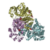
- Structure visualization
| Structure viewer | Molecule: MolmilJmol/JSmol |
|---|
- Downloads & links
-Download
| PDBx/mmCIF format | 6jl6.cif.gz | 362 KB | Display | PDBx/mmCIF format |
|---|---|---|---|---|
| PDB format | pdb6jl6.ent.gz | 296.9 KB | Display | PDB format |
| PDBx/mmJSON format | 6jl6.json.gz | Tree view | PDBx/mmJSON format | |
| Others |  Other downloads Other downloads |
-Validation report
| Arichive directory | https://data.pdbj.org/pub/pdb/validation_reports/jl/6jl6ftp://data.pdbj.org/pub/pdb/validation_reports/jl/6jl6 | HTTPS FTP |
|---|
-Related structure data
| Related structure data |  6jkqSC  6jkrC  6jksC  6jktC  6jl4C  6jl5C S: Starting model for refinement C: citing same article ( |
|---|---|
| Similar structure data |


-Links
 PDBj
PDBj

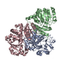
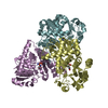
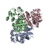
- Assembly
| Deposited unit | 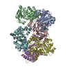
| ||||||||
|---|---|---|---|---|---|---|---|---|---|
| 1 | 
| ||||||||
| 2 | 
| ||||||||
| Unit cell |
|
-Components
| #1: Protein | Mass: 36081.766 Da / Num. of mol.: 6 Source method: isolated from a genetically manipulated source Source: (gene. exp.)  #2: Chemical | ChemComp-ASP / |   Type: L-peptide linking / Mass: 133.103 Da / Num. of mol.: 1 / Source method: obtained synthetically / Formula: C4H7NO4 Type: L-peptide linking / Mass: 133.103 Da / Num. of mol.: 1 / Source method: obtained synthetically / Formula: C4H7NO4#3: Chemical | ChemComp-PO4 / |   Mass: 94.971 Da / Num. of mol.: 1 / Source method: obtained synthetically / Formula: PO4 Mass: 94.971 Da / Num. of mol.: 1 / Source method: obtained synthetically / Formula: PO4#4: Water | ChemComp-HOH / |  Mass: 18.015 Da / Num. of mol.: 434 / Source method: isolated from a natural source / Formula: H2O Mass: 18.015 Da / Num. of mol.: 434 / Source method: isolated from a natural source / Formula: H2O |
|---|
-Experimental details
-Experiment
| Experiment | Method: X-RAY DIFFRACTION / Number of used crystals: 1 |
|---|
- Sample preparation
| Crystal | Density Matthews: 2.5 Å3/Da / Density % sol: 50.9 % |
|---|---|
| Crystal grow | Temperature: 293 K / Method: vapor diffusion, hanging drop / pH: 4.6 Details: 10% PEG 3350, 0.1 M acetate buffer, pH 4.6, 0.2 M ammonium acetate, 0.01 M cobalt chloride, 3% glycerol. |
-Data collection
| Diffraction | Mean temperature: 100 K / Serial crystal experiment: N |
|---|---|
| Diffraction source | Source: SYNCHROTRON / Site: SPring-8  / Beamline: BL44XU / Wavelength: 0.9 Å / Beamline: BL44XU / Wavelength: 0.9 Å |
| Detector | Type: MARMOSAIC 300 mm CCD / Detector: CCD / Date: Jul 20, 2017 |
| Radiation | Monochromator: Si(111) / Protocol: SINGLE WAVELENGTH / Monochromatic (M) / Laue (L): M / Scattering type: x-ray |
| Radiation wavelength | Wavelength: 0.9 Å / Relative weight: 1 |
| Reflection | Resolution: 2→50 Å / Num. obs: 143565 / % possible obs: 99.9 % / Redundancy: 3.6 % / Rmerge(I) obs: 0.119 / Net I/σ(I): 9.1 |
| Reflection shell | Resolution: 2→2.03 Å / Redundancy: 3.4 % / Rmerge(I) obs: 0.56 / Mean I/σ(I) obs: 2.5 / Num. unique obs: 7167 / % possible all: 100 |
- Processing
| Software |
| ||||||||||||||||||||||||||||||||||||||||||||||||||||||||||||||||||||||||||||||||||||||||||||||||||||||||||||||||||||||||||||||||||||||||||||||||||||||||||||||||||||||||||||||||||||||
|---|---|---|---|---|---|---|---|---|---|---|---|---|---|---|---|---|---|---|---|---|---|---|---|---|---|---|---|---|---|---|---|---|---|---|---|---|---|---|---|---|---|---|---|---|---|---|---|---|---|---|---|---|---|---|---|---|---|---|---|---|---|---|---|---|---|---|---|---|---|---|---|---|---|---|---|---|---|---|---|---|---|---|---|---|---|---|---|---|---|---|---|---|---|---|---|---|---|---|---|---|---|---|---|---|---|---|---|---|---|---|---|---|---|---|---|---|---|---|---|---|---|---|---|---|---|---|---|---|---|---|---|---|---|---|---|---|---|---|---|---|---|---|---|---|---|---|---|---|---|---|---|---|---|---|---|---|---|---|---|---|---|---|---|---|---|---|---|---|---|---|---|---|---|---|---|---|---|---|---|---|---|---|---|
| Refinement | Method to determine structure: MOLECULAR REPLACEMENT Starting model: 6JKQ Resolution: 2→20 Å / Cor.coef. Fo:Fc: 0.95 / Cor.coef. Fo:Fc free: 0.929 / SU B: 4.133 / SU ML: 0.114 / Cross valid method: THROUGHOUT / ESU R: 0.179 / ESU R Free: 0.158 / Details: HYDROGENS HAVE BEEN ADDED IN THE RIDING POSITIONS
| ||||||||||||||||||||||||||||||||||||||||||||||||||||||||||||||||||||||||||||||||||||||||||||||||||||||||||||||||||||||||||||||||||||||||||||||||||||||||||||||||||||||||||||||||||||||
| Solvent computation | Ion probe radii: 0.8 Å / Shrinkage radii: 0.8 Å / VDW probe radii: 1.2 Å | ||||||||||||||||||||||||||||||||||||||||||||||||||||||||||||||||||||||||||||||||||||||||||||||||||||||||||||||||||||||||||||||||||||||||||||||||||||||||||||||||||||||||||||||||||||||
| Displacement parameters | Biso mean: 27.803 Å2
| ||||||||||||||||||||||||||||||||||||||||||||||||||||||||||||||||||||||||||||||||||||||||||||||||||||||||||||||||||||||||||||||||||||||||||||||||||||||||||||||||||||||||||||||||||||||
| Refinement step | Cycle: 1 / Resolution: 2→20 Å
| ||||||||||||||||||||||||||||||||||||||||||||||||||||||||||||||||||||||||||||||||||||||||||||||||||||||||||||||||||||||||||||||||||||||||||||||||||||||||||||||||||||||||||||||||||||||
| Refine LS restraints |
|