 Movie
Movie Controller

[English] 日本語
 Yorodumi
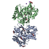
- PDB-2j7t: Crystal structure of human serine threonine kinase-10 bound to SU11274 -
+ Open data

- Basic information
| Entry | Database: PDB / ID: 2j7t | ||||||
|---|---|---|---|---|---|---|---|
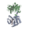
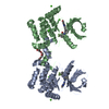
| Title | Crystal structure of human serine threonine kinase-10 bound to SU11274 | ||||||
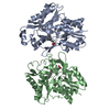
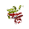
 Components Components | SERINE/THREONINE-PROTEIN KINASE 10 | ||||||
 Keywords Keywords | TRANSFERASE / KINASE / ATP-BINDING / CELL CYCLE PROGRESSION / PHOSPHORYLATION / DISEASE MUTATION / NUCLEOTIDE- BINDING / LYMPHOCYTE ORIENTED KINASE (LOK) / SERINE/THREONINE- PROTEIN KINASE / SERINE/THREONINE KINASE (STK10A) | ||||||
| Function / homology |  Function and homology information Function and homology informationlymphocyte aggregation / regulation of lymphocyte migration / RHOB GTPase cycle / RHOC GTPase cycle / RHOA GTPase cycle / specific granule membrane / protein autophosphorylation / protein phosphorylation / non-specific serine/threonine protein kinase / intracellular signal transduction ...lymphocyte aggregation / regulation of lymphocyte migration / RHOB GTPase cycle / RHOC GTPase cycle / RHOA GTPase cycle / specific granule membrane / protein autophosphorylation / protein phosphorylation / non-specific serine/threonine protein kinase / intracellular signal transduction / nuclear body / protein serine kinase activity / protein serine/threonine kinase activity / Neutrophil degranulation / protein homodimerization activity / extracellular exosome / nucleoplasm / ATP binding / identical protein binding / plasma membrane / cytoplasm / cytosol Similarity search - Function | ||||||
| Biological species |  HOMO SAPIENS (human) HOMO SAPIENS (human) | ||||||
| Method |  X-RAY DIFFRACTION / SYNCHROTRON / MOLECULAR REPLACEMENT / Resolution: 2 Å X-RAY DIFFRACTION / SYNCHROTRON / MOLECULAR REPLACEMENT / Resolution: 2 Å | ||||||
 Authors Authors | Pike, A.C.W. / Rellos, P. / Fedorov, O. / Das, S. / Debreczeni, J. / Sobott, F. / Watt, S. / Savitsky, P. / Eswaran, J. / Turnbull, A.P. ...Pike, A.C.W. / Rellos, P. / Fedorov, O. / Das, S. / Debreczeni, J. / Sobott, F. / Watt, S. / Savitsky, P. / Eswaran, J. / Turnbull, A.P. / Papagrigoriou, E. / Ugochukwa, E. / Gorrec, F. / Umeano, C.C. / von Delft, F. / Arrowsmith, C.H. / Edwards, A. / Weigelt, J. / Sundstrom, M. / Knapp, S. | ||||||
 Citation Citation | Journal: Embo J. / Year: 2008 Title: Activation Segment Dimerization: A Mechanism for Kinase Autophosphorylation of Non-Consensus Sites. Authors: Pike, A.C.W. / Rellos, P. / Niesen, F.H. / Turnbull, A. / Oliver, A.W. / Parker, S.A. / Turk, B.E. / Pearl, L.H. / Knapp, S. | ||||||
| History |
|
- Structure visualization
| Structure viewer | Molecule: MolmilJmol/JSmol |
|---|
- Downloads & links
-Download
| PDBx/mmCIF format | 2j7t.cif.gz | 75.8 KB | Display | PDBx/mmCIF format |
|---|---|---|---|---|
| PDB format | pdb2j7t.ent.gz | 54.4 KB | Display | PDB format |
| PDBx/mmJSON format | 2j7t.json.gz | Tree view | PDBx/mmJSON format | |
| Others |  Other downloads Other downloads |
-Validation report
| Arichive directory | https://data.pdbj.org/pub/pdb/validation_reports/j7/2j7tftp://data.pdbj.org/pub/pdb/validation_reports/j7/2j7t | HTTPS FTP |
|---|
-Related structure data
| Related structure data |  2j51SC 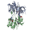 2j90C  2jflC 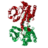 2jfmC  2uv2C S: Starting model for refinement C: citing same article ( |
|---|---|
| Similar structure data |


-Links
 PDBj
PDBj
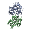
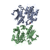
- Assembly
| Deposited unit | 
| ||||||||
|---|---|---|---|---|---|---|---|---|---|
| 1 | 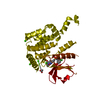
| ||||||||
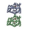
| Unit cell |
|
-Components
| #1: Protein | Mass: 34298.605 Da / Num. of mol.: 1 / Fragment: KINASE DOMAIN, RESIDUES 18-317 Source method: isolated from a genetically manipulated source Source: (gene. exp.) HOMO SAPIENS (human) / Plasmid: PNIC28-BSA4 / Production host:  References: UniProt: O94804, non-specific serine/threonine protein kinase | ||||||||||
|---|---|---|---|---|---|---|---|---|---|---|---|
| #2: Chemical | ChemComp-CA /   Mass: 40.078 Da / Num. of mol.: 7 / Source method: obtained synthetically / Formula: Ca Mass: 40.078 Da / Num. of mol.: 7 / Source method: obtained synthetically / Formula: Ca#3: Chemical | ChemComp-ACT / |   Mass: 59.044 Da / Num. of mol.: 1 / Source method: obtained synthetically / Formula: C2H3O2 Mass: 59.044 Da / Num. of mol.: 1 / Source method: obtained synthetically / Formula: C2H3O2#4: Chemical | ChemComp-274 / ( | 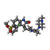  Mass: 568.087 Da / Num. of mol.: 1 / Source method: obtained synthetically / Formula: C28H30ClN5O4S Mass: 568.087 Da / Num. of mol.: 1 / Source method: obtained synthetically / Formula: C28H30ClN5O4S#5: Water | ChemComp-HOH / |  Mass: 18.015 Da / Num. of mol.: 130 / Source method: isolated from a natural source / Formula: H2O Mass: 18.015 Da / Num. of mol.: 130 / Source method: isolated from a natural source / Formula: H2OHas protein modification | Y | Sequence details | SEQUENCE DISCREPANC | |
-Experimental details
-Experiment
| Experiment | Method: X-RAY DIFFRACTION / Number of used crystals: 1 |
|---|
- Sample preparation
| Crystal | Density Matthews: 2.71 Å3/Da / Density % sol: 54.6 % |
|---|---|
| Crystal grow | pH: 6.5 Details: 45% PEG300, 0.24M CALCIUM ACETATE, 0.1M SODIUM CACODYLATE PH6.5, pH 6.50 |
-Data collection
| Diffraction | Mean temperature: 100 K |
|---|---|
| Diffraction source | Source: SYNCHROTRON / Site: SLS  / Beamline: X10SA / Wavelength: 0.999 / Beamline: X10SA / Wavelength: 0.999 |
| Detector | Type: MARRESEARCH / Detector: CCD / Date: Sep 23, 2006 |
| Radiation | Protocol: SINGLE WAVELENGTH / Monochromatic (M) / Laue (L): M / Scattering type: x-ray |
| Radiation wavelength | Wavelength: 0.999 Å / Relative weight: 1 |
| Reflection | Resolution: 2→56.52 Å / Num. obs: 25648 / % possible obs: 99.9 % / Observed criterion σ(I): 0 / Redundancy: 5.3 % / Biso Wilson estimate: 28.7 Å2 / Rmerge(I) obs: 0.09 / Net I/σ(I): 11.4 |
| Reflection shell | Resolution: 2→2.11 Å / Redundancy: 5.3 % / Rmerge(I) obs: 0.82 / Mean I/σ(I) obs: 2.3 / % possible all: 99.8 |
- Processing
| Software |
| ||||||||||||||||||||||||||||||||||||||||||||||||||||||||||||||||||||||||||||||||||||||||||||||||||||||||||||||||||||||||||||||||||||||||||||||||||||||||||||||||||||||||||||||||||||||
|---|---|---|---|---|---|---|---|---|---|---|---|---|---|---|---|---|---|---|---|---|---|---|---|---|---|---|---|---|---|---|---|---|---|---|---|---|---|---|---|---|---|---|---|---|---|---|---|---|---|---|---|---|---|---|---|---|---|---|---|---|---|---|---|---|---|---|---|---|---|---|---|---|---|---|---|---|---|---|---|---|---|---|---|---|---|---|---|---|---|---|---|---|---|---|---|---|---|---|---|---|---|---|---|---|---|---|---|---|---|---|---|---|---|---|---|---|---|---|---|---|---|---|---|---|---|---|---|---|---|---|---|---|---|---|---|---|---|---|---|---|---|---|---|---|---|---|---|---|---|---|---|---|---|---|---|---|---|---|---|---|---|---|---|---|---|---|---|---|---|---|---|---|---|---|---|---|---|---|---|---|---|---|---|
| Refinement | Method to determine structure: MOLECULAR REPLACEMENT Starting model: PDB ENTRY 2J51 Resolution: 2→55 Å / Cor.coef. Fo:Fc: 0.946 / Cor.coef. Fo:Fc free: 0.928 / SU B: 7.399 / SU ML: 0.11 / TLS residual ADP flag: LIKELY RESIDUAL / Cross valid method: THROUGHOUT / ESU R: 0.164 / ESU R Free: 0.148 / Stereochemistry target values: MAXIMUM LIKELIHOOD / Details: HYDROGENS HAVE BEEN ADDED IN THE RIDING POSITIONS.
| ||||||||||||||||||||||||||||||||||||||||||||||||||||||||||||||||||||||||||||||||||||||||||||||||||||||||||||||||||||||||||||||||||||||||||||||||||||||||||||||||||||||||||||||||||||||
| Solvent computation | Ion probe radii: 0.8 Å / Shrinkage radii: 0.8 Å / VDW probe radii: 1.4 Å / Solvent model: MASK | ||||||||||||||||||||||||||||||||||||||||||||||||||||||||||||||||||||||||||||||||||||||||||||||||||||||||||||||||||||||||||||||||||||||||||||||||||||||||||||||||||||||||||||||||||||||
| Displacement parameters | Biso mean: 32.29 Å2
| ||||||||||||||||||||||||||||||||||||||||||||||||||||||||||||||||||||||||||||||||||||||||||||||||||||||||||||||||||||||||||||||||||||||||||||||||||||||||||||||||||||||||||||||||||||||
| Refinement step | Cycle: LAST / Resolution: 2→55 Å
| ||||||||||||||||||||||||||||||||||||||||||||||||||||||||||||||||||||||||||||||||||||||||||||||||||||||||||||||||||||||||||||||||||||||||||||||||||||||||||||||||||||||||||||||||||||||
| Refine LS restraints |
|