 Movie
Movie Controller


+ Open data

- Basic information
| Entry | Database: PDB / ID: 4rnx | ||||||
|---|---|---|---|---|---|---|---|
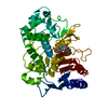
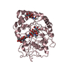
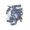
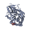
| Title | K154 Circular Permutation of Old Yellow Enzyme | ||||||
 Components Components | NADPH dehydrogenase 1 | ||||||
 Keywords Keywords | OXIDOREDUCTASE / CIRCULAR PERMUTATION / CATALYSIS / OLD YELLOW ENZYME / FLAVIN COFACTOR | ||||||
| Function / homology |  Function and homology information Function and homology informationNADPH dehydrogenase / NADPH dehydrogenase activity / FMN binding / apoptotic process Similarity search - Function | ||||||
| Biological species |  | ||||||
| Method |  X-RAY DIFFRACTION / SYNCHROTRON / MOLECULAR REPLACEMENT / Resolution: 1.25 Å X-RAY DIFFRACTION / SYNCHROTRON / MOLECULAR REPLACEMENT / Resolution: 1.25 Å | ||||||
 Authors Authors | Horton, J.R. / Daugherty, A.B. / Cheng, X. / Lutz, S. | ||||||
 Citation Citation | Journal: ACS Catal / Year: 2015 Title: STRUCTURAL AND FUNCTIONAL CONSEQUENCES OF CIRCULAR PERMUTATION ON THE ACTIVE SITE OF OLD YELLOW ENZYME. Authors: Daugherty, A.B. / Horton, J.R. / Cheng, X. / Lutz, S. | ||||||
| History |
|
- Structure visualization
| Structure viewer | Molecule: MolmilJmol/JSmol |
|---|
- Downloads & links
-Download
| PDBx/mmCIF format | 4rnx.cif.gz | 342 KB | Display | PDBx/mmCIF format |
|---|---|---|---|---|
| PDB format | pdb4rnx.ent.gz | 274.8 KB | Display | PDB format |
| PDBx/mmJSON format | 4rnx.json.gz | Tree view | PDBx/mmJSON format | |
| Others |  Other downloads Other downloads |
-Validation report
| Arichive directory | https://data.pdbj.org/pub/pdb/validation_reports/rn/4rnxftp://data.pdbj.org/pub/pdb/validation_reports/rn/4rnx | HTTPS FTP |
|---|
-Related structure data
| Related structure data |  4rnuC  4rnvC  4rnwC  1oyaS C: citing same article ( S: Starting model for refinement |
|---|---|
| Similar structure data |


-Links
 PDBj
PDBj
- Assembly
| Deposited unit | 
| ||||||||
|---|---|---|---|---|---|---|---|---|---|
| 1 | 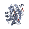
| ||||||||
| 2 | 
| ||||||||
| Unit cell |
|
-Components
| #1: Protein | Mass: 44943.445 Da / Num. of mol.: 2 / Fragment: UNP residues 154-397, 2-153 Source method: isolated from a genetically manipulated source Source: (gene. exp.) Gene: OYE1 / Plasmid: pET-14b / Production host:  #2: Chemical |   Mass: 456.344 Da / Num. of mol.: 2 / Source method: obtained synthetically / Formula: C17H21N4O9P Mass: 456.344 Da / Num. of mol.: 2 / Source method: obtained synthetically / Formula: C17H21N4O9P#3: Chemical | ChemComp-EDO /   Mass: 62.068 Da / Num. of mol.: 6 / Source method: obtained synthetically / Formula: C2H6O2 Mass: 62.068 Da / Num. of mol.: 6 / Source method: obtained synthetically / Formula: C2H6O2#4: Water | ChemComp-HOH / |  Mass: 18.015 Da / Num. of mol.: 1121 / Source method: isolated from a natural source / Formula: H2O Mass: 18.015 Da / Num. of mol.: 1121 / Source method: isolated from a natural source / Formula: H2O |
|---|
-Experimental details
-Experiment
| Experiment | Method: X-RAY DIFFRACTION / Number of used crystals: 1 |
|---|
- Sample preparation
| Crystal | Density Matthews: 2.16 Å3/Da / Density % sol: 42.95 % |
|---|---|
| Crystal grow | Temperature: 289 K / Method: vapor diffusion, hanging drop / pH: 5 Details: 30% PEG 2KMME, 0.1M Potassium Thiocyanate, pH 5.0, VAPOR DIFFUSION, HANGING DROP, temperature 289K |
-Data collection
| Diffraction | Mean temperature: 100 K |
|---|---|
| Diffraction source | Source: SYNCHROTRON / Site: APS  / Beamline: 22-BM / Wavelength: 1 Å / Beamline: 22-BM / Wavelength: 1 Å |
| Detector | Type: MARMOSAIC 300 mm CCD / Detector: CCD / Date: Aug 16, 2013 |
| Radiation | Monochromator: Si(111) / Protocol: SINGLE WAVELENGTH / Monochromatic (M) / Laue (L): M / Scattering type: x-ray |
| Radiation wavelength | Wavelength: 1 Å / Relative weight: 1 |
| Reflection | Resolution: 1.25→27.11 Å / Num. all: 207000 / Num. obs: 196866 / % possible obs: 95.1 % / Observed criterion σ(F): -3 / Observed criterion σ(I): -3 / Redundancy: 4.2 % / Biso Wilson estimate: 10.2 Å2 / Rmerge(I) obs: 0.079 / Net I/σ(I): 17 |
| Reflection shell | Resolution: 1.25→1.29 Å / Redundancy: 4 % / Rmerge(I) obs: 0.681 / Mean I/σ(I) obs: 1.9 / Num. unique all: 19029 / % possible all: 92 |
- Processing
| Software |
| |||||||||||||||||||||||||||||||||||||||||||||||||||||||||||||||||||||||||||||||||||||||||||||||||||||||||
|---|---|---|---|---|---|---|---|---|---|---|---|---|---|---|---|---|---|---|---|---|---|---|---|---|---|---|---|---|---|---|---|---|---|---|---|---|---|---|---|---|---|---|---|---|---|---|---|---|---|---|---|---|---|---|---|---|---|---|---|---|---|---|---|---|---|---|---|---|---|---|---|---|---|---|---|---|---|---|---|---|---|---|---|---|---|---|---|---|---|---|---|---|---|---|---|---|---|---|---|---|---|---|---|---|---|---|
| Refinement | Method to determine structure: MOLECULAR REPLACEMENT Starting model: PDB entry 1OYA Resolution: 1.25→27.11 Å / SU ML: 0.11 / Cross valid method: THROUGHOUT / σ(F): 1.96 / Phase error: 16.23 / Stereochemistry target values: ML
| |||||||||||||||||||||||||||||||||||||||||||||||||||||||||||||||||||||||||||||||||||||||||||||||||||||||||
| Solvent computation | Shrinkage radii: 0.9 Å / VDW probe radii: 1.11 Å / Solvent model: FLAT BULK SOLVENT MODEL | |||||||||||||||||||||||||||||||||||||||||||||||||||||||||||||||||||||||||||||||||||||||||||||||||||||||||
| Refinement step | Cycle: LAST / Resolution: 1.25→27.11 Å
| |||||||||||||||||||||||||||||||||||||||||||||||||||||||||||||||||||||||||||||||||||||||||||||||||||||||||
| Refine LS restraints |
| |||||||||||||||||||||||||||||||||||||||||||||||||||||||||||||||||||||||||||||||||||||||||||||||||||||||||
| LS refinement shell |
|
