 Movie
Movie Controller

[English] 日本語
 Yorodumi
- PDB-1nwt: Crystal structure of human cartilage gp39 (HC-gp39) in complex wi... -
+ Open data

- Basic information
| Entry | Database: PDB / ID: 1nwt | |||||||||
|---|---|---|---|---|---|---|---|---|---|---|
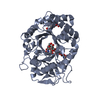
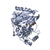
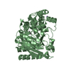
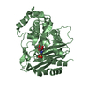
| Title | Crystal structure of human cartilage gp39 (HC-gp39) in complex with chitopentaose | |||||||||
 Components Components | Chitinase-3 like protein 1 | |||||||||
 Keywords Keywords | SIGNALING PROTEIN / chitinase-like protein / rheumatoid arthritis / chitin / N-acetylglucosamine | |||||||||
| Function / homology |  Function and homology information Function and homology informationresponse to interleukin-6 / activation of NF-kappaB-inducing kinase activity / cartilage development / positive regulation of peptidyl-threonine phosphorylation / chitin catabolic process / extracellular matrix structural constituent / chitin binding / response to tumor necrosis factor / response to mechanical stimulus / lung development ...response to interleukin-6 / activation of NF-kappaB-inducing kinase activity / cartilage development / positive regulation of peptidyl-threonine phosphorylation / chitin catabolic process / extracellular matrix structural constituent / chitin binding / response to tumor necrosis factor / response to mechanical stimulus / lung development / response to interleukin-1 / positive regulation of interleukin-8 production / specific granule lumen / cellular response to tumor necrosis factor / positive regulation of angiogenesis / carbohydrate binding / extracellular matrix / carbohydrate metabolic process / positive regulation of ERK1 and ERK2 cascade / positive regulation of phosphatidylinositol 3-kinase/protein kinase B signal transduction / inflammatory response / apoptotic process / Neutrophil degranulation / perinuclear region of cytoplasm / endoplasmic reticulum / : / extracellular exosome / extracellular region / cytoplasm Similarity search - Function | |||||||||
| Biological species |  Homo sapiens (human) Homo sapiens (human) | |||||||||
| Method |  X-RAY DIFFRACTION / MOLECULAR REPLACEMENT / Resolution: 2.5 Å X-RAY DIFFRACTION / MOLECULAR REPLACEMENT / Resolution: 2.5 Å | |||||||||
 Authors Authors | Fusetti, F. / Pijning, T. / Kalk, K.H. / Bos, E. / Dijkstra, B.W. | |||||||||
 Citation Citation | Journal: J.Biol.Chem. / Year: 2003 Title: Crystal structure and carbohydrate-binding properties of the human cartilage glycoprotein-39 Authors: Fusetti, F. / Pijning, T. / Kalk, K.H. / Bos, E. / Dijkstra, B.W. | |||||||||
| History |
| |||||||||
| Remark 700 | SHEET DETERMINATION METHOD: DSSP THE SHEET PRESENTED AS A1 ON SHEET RECORDS BELOW IS ACTUALLY AN ... SHEET DETERMINATION METHOD: DSSP THE SHEET PRESENTED AS A1 ON SHEET RECORDS BELOW IS ACTUALLY AN EIGHT-STRANDED ALPHA/BETA-BARREL. THIS IS REPRESENTED BY A NINE-STRANDED SHEET IN WHICH THE FIRST AND LAST STRANDS ARE IDENTICAL. THE SHEET PRESENTED AS B1 ON SHEET RECORDS BELOW IS ACTUALLY AN EIGHT-STRANDED ALPHA/BETA-BARREL. THIS IS REPRESENTED BY A NINE-STRANDED SHEET IN WHICH THE FIRST AND LAST STRANDS ARE IDENTICAL. THE SHEET PRESENTED AS C1 ON SHEET RECORDS BELOW IS ACTUALLY AN EIGHT-STRANDED ALPHA/BETA-BARREL. THIS IS REPRESENTED BY A NINE-STRANDED SHEET IN WHICH THE FIRST AND LAST STRANDS ARE IDENTICAL. THE SHEET PRESENTED AS D1 ON SHEET RECORDS BELOW IS ACTUALLY AN EIGHT-STRANDED ALPHA/BETA-BARREL. THIS IS REPRESENTED BY A NINE-STRANDED SHEET IN WHICH THE FIRST AND LAST STRANDS ARE IDENTICAL. SHEETS A3A AND A3B ARE IDENTICAL EXCEPT FOR THE FIRST STRAND. THIS STRAND IS SPLIT INTO TWO DISTINCT AMINO ACID RUNS. THE BULGE OF 11 AMINO ACIDS (RESIDUES 282-292) INCLUDES A HYDROGEN-BONDED TURN (RESIDUES 286-292). SHEETS B3A AND B3B ARE IDENTICAL EXCEPT FOR THE FIRST STRAND. THIS STRAND IS SPLIT INTO TWO DISTINCT AMINO ACID RUNS. THE BULGE OF 11 AMINO ACIDS (RESIDUES 282-292) INCLUDES A HYDROGEN-BONDED TURN (RESIDUES 286-292). SHEETS C3A AND C3B ARE IDENTICAL EXCEPT FOR THE FIRST STRAND. THIS STRAND IS SPLIT INTO TWO DISTINCT AMINO ACID RUNS. THE BULGE OF 11 AMINO ACIDS (RESIDUES 282-292) INCLUDES A HYDROGEN-BONDED TURN (RESIDUES 286-292). SHEETS D3A AND D3B ARE IDENTICAL EXCEPT FOR THE FIRST STRAND. THIS STRAND IS SPLIT INTO TWO DISTINCT AMINO ACID RUNS. THE BULGE OF 11 AMINO ACIDS (RESIDUES 282-292) INCLUDES A HYDROGEN-BONDED TURN (RESIDUES 286-292). |
- Structure visualization
| Structure viewer | Molecule: MolmilJmol/JSmol |
|---|
- Downloads & links
-Download
| PDBx/mmCIF format | 1nwt.cif.gz | 306.5 KB | Display | PDBx/mmCIF format |
|---|---|---|---|---|
| PDB format | pdb1nwt.ent.gz | 251.1 KB | Display | PDB format |
| PDBx/mmJSON format | 1nwt.json.gz | Tree view | PDBx/mmJSON format | |
| Others |  Other downloads Other downloads |
-Validation report
| Arichive directory | https://data.pdbj.org/pub/pdb/validation_reports/nw/1nwtftp://data.pdbj.org/pub/pdb/validation_reports/nw/1nwt | HTTPS FTP |
|---|
-Related structure data


-Links
 PDBj
PDBj- Assembly
| Deposited unit | 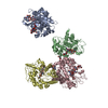
| ||||||||
|---|---|---|---|---|---|---|---|---|---|
| 1 | 
| ||||||||
| 2 | 
| ||||||||
| 3 | 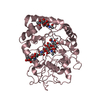
| ||||||||
| 4 | 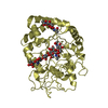
| ||||||||
| Unit cell |
|
-Components
| #1: Protein | Mass: 40536.758 Da / Num. of mol.: 4 Source method: isolated from a genetically manipulated source Source: (gene. exp.) Homo sapiens (human) / Cell: synovial cells, articular chondrocytes / Cell (production host): mammalian cho cells / Production host:   Cricetulus griseus (Chinese hamster) / References: UniProt: P36222 Cricetulus griseus (Chinese hamster) / References: UniProt: P36222#2: Polysaccharide | 2-acetamido-2-deoxy-beta-D-glucopyranose-(1-4)-2-acetamido-2-deoxy-beta-D-glucopyranose Source method: isolated from a genetically manipulated source #3: Polysaccharide | Source method: isolated from a genetically manipulated source #4: Polysaccharide | 2-acetamido-2-deoxy-beta-D-glucopyranose-(1-4)-2-acetamido-2-deoxy-alpha-D-glucopyranose-(1-4)-2- ...2-acetamido-2-deoxy-beta-D-glucopyranose-(1-4)-2-acetamido-2-deoxy-alpha-D-glucopyranose-(1-4)-2-acetamido-2-deoxy-beta-D-glucopyranose-(1-4)-2-acetamido-2-deoxy-beta-D-glucopyranose-(1-4)-2-acetamido-2-deoxy-alpha-D-glucopyranose | Source method: isolated from a genetically manipulated source #5: Water | ChemComp-HOH / |  Mass: 18.015 Da / Num. of mol.: 470 / Source method: isolated from a natural source / Formula: H2O Mass: 18.015 Da / Num. of mol.: 470 / Source method: isolated from a natural source / Formula: H2OHas protein modification | Y | |
|---|
-Experimental details
-Experiment
| Experiment | Method: X-RAY DIFFRACTION |
|---|
- Sample preparation
| Crystal | Density Matthews: 2.6 Å3/Da / Density % sol: 50.23 % | |||||||||||||||||||||||||||||||||||||||||||||||||
|---|---|---|---|---|---|---|---|---|---|---|---|---|---|---|---|---|---|---|---|---|---|---|---|---|---|---|---|---|---|---|---|---|---|---|---|---|---|---|---|---|---|---|---|---|---|---|---|---|---|---|
| Crystal grow | pH: 5.1 / Details: pH 5.1 | |||||||||||||||||||||||||||||||||||||||||||||||||
| Crystal grow | *PLUS pH: 7.2 / Method: vapor diffusion, hanging drop | |||||||||||||||||||||||||||||||||||||||||||||||||
| Components of the solutions | *PLUS
|
-Data collection
| Diffraction | Mean temperature: 120 K |
|---|---|
| Diffraction source | Source: ROTATING ANODE / Type: OTHER |
| Detector | Type: MAC Science DIP-2030 / Detector: IMAGE PLATE |
| Radiation | Protocol: SINGLE WAVELENGTH / Monochromatic (M) / Laue (L): M / Scattering type: x-ray |
| Radiation wavelength | Relative weight: 1 |
| Reflection | Resolution: 2.5→29.92 Å / Num. obs: 59975 / % possible obs: 97.9 % / Observed criterion σ(I): 0 / Redundancy: 3.8 % / Biso Wilson estimate: 45.5 Å2 / Rsym value: 0.114 / Net I/σ(I): 10.1 |
| Reflection shell | Highest resolution: 2.5 Å / Mean I/σ(I) obs: 2 / Rsym value: 0.626 / % possible all: 96.1 |
| Reflection | *PLUS Highest resolution: 2.5 Å / Lowest resolution: 30 Å / Num. measured all: 227637 / Rmerge(I) obs: 0.114 |
| Reflection shell | *PLUS % possible obs: 96.1 % / Rmerge(I) obs: 0.626 |
- Processing
| Software |
| ||||||||||||||||||||||||||||||||||||||||||||||||||||||||||||||||||||||||||||||||
|---|---|---|---|---|---|---|---|---|---|---|---|---|---|---|---|---|---|---|---|---|---|---|---|---|---|---|---|---|---|---|---|---|---|---|---|---|---|---|---|---|---|---|---|---|---|---|---|---|---|---|---|---|---|---|---|---|---|---|---|---|---|---|---|---|---|---|---|---|---|---|---|---|---|---|---|---|---|---|---|---|---|
| Refinement | Method to determine structure: MOLECULAR REPLACEMENT / Resolution: 2.5→29.92 Å / Rfactor Rfree error: 0.003 / Data cutoff high absF: 2447799.71 / Data cutoff high rms absF: 2447799.71 / Data cutoff low absF: 0 / Isotropic thermal model: RESTRAINED / Cross valid method: THROUGHOUT / σ(F): 0 Details: NON-CRYSTALLOGRAPHIC SYMMETRY RESTRAINTS WERE IMPOSED ON THE FOLLOWING RESIDUE GROUPS IN ALL FOUR MOLECULES: GROUP 1: RESIDUES 22 TO 208, GROUP 2: RESIDUES 214 TO 226, GROUP 3: RESIDUES 235 ...Details: NON-CRYSTALLOGRAPHIC SYMMETRY RESTRAINTS WERE IMPOSED ON THE FOLLOWING RESIDUE GROUPS IN ALL FOUR MOLECULES: GROUP 1: RESIDUES 22 TO 208, GROUP 2: RESIDUES 214 TO 226, GROUP 3: RESIDUES 235 TO 242, GROUP 4: RESIDUES 249 to 383
| ||||||||||||||||||||||||||||||||||||||||||||||||||||||||||||||||||||||||||||||||
| Solvent computation | Solvent model: FLAT MODEL / Bsol: 40.53 Å2 / ksol: 0.35 e/Å3 | ||||||||||||||||||||||||||||||||||||||||||||||||||||||||||||||||||||||||||||||||
| Displacement parameters | Biso mean: 36.9 Å2
| ||||||||||||||||||||||||||||||||||||||||||||||||||||||||||||||||||||||||||||||||
| Refine analyze |
| ||||||||||||||||||||||||||||||||||||||||||||||||||||||||||||||||||||||||||||||||
| Refinement step | Cycle: LAST / Resolution: 2.5→29.92 Å
| ||||||||||||||||||||||||||||||||||||||||||||||||||||||||||||||||||||||||||||||||
| Refine LS restraints |
| ||||||||||||||||||||||||||||||||||||||||||||||||||||||||||||||||||||||||||||||||
| LS refinement shell | Resolution: 2.5→2.66 Å / Rfactor Rfree error: 0.013 / Total num. of bins used: 6
| ||||||||||||||||||||||||||||||||||||||||||||||||||||||||||||||||||||||||||||||||
| Xplor file |
| ||||||||||||||||||||||||||||||||||||||||||||||||||||||||||||||||||||||||||||||||
| Refinement | *PLUS Highest resolution: 2.5 Å / Lowest resolution: 30 Å / % reflection Rfree: 10 % / Rfactor Rwork: 0.217 | ||||||||||||||||||||||||||||||||||||||||||||||||||||||||||||||||||||||||||||||||
| Solvent computation | *PLUS | ||||||||||||||||||||||||||||||||||||||||||||||||||||||||||||||||||||||||||||||||
| Displacement parameters | *PLUS | ||||||||||||||||||||||||||||||||||||||||||||||||||||||||||||||||||||||||||||||||
| Refine LS restraints | *PLUS
|
