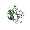
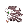
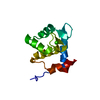
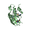
BIOMOLECULE: 1, 2 SEE REMARK 350 FOR THE AUTHOR PROVIDED AND PROGRAM GENERATED ASSEMBLY ... BIOMOLECULE: 1, 2 SEE REMARK 350 FOR THE AUTHOR PROVIDED AND PROGRAM GENERATED ASSEMBLY INFORMATION FOR THE STRUCTURE IN THIS ENTRY. THE REMARK MAY ALSO PROVIDE INFORMATION ON BURIED SURFACE AREA. SIZE EXCLUSION CHROMATOGRAPHY WITH STATIC LIGHT SCATTERING SUPPORTS THE ASSIGNMENT OF A MONOMER AS A SIGNIFICANT OLIGOMERIZATION STATE IN SOLUTION.
Remark 999
SEQUENCE THE CONSTRUCT WAS EXPRESSED WITH A PURIFICATION TAG MGSDKIHHHHHHENLYFQG.
Type: MARMOSAIC 300 mm CCD / Detector: CCD / Date: Aug 19, 2007 / Details: Adjustable focusing mirrors in K-B geometry
Radiation
Monochromator: Si(111) Double crystal / Protocol: SINGLE WAVELENGTH / Monochromatic (M) / Laue (L): M / Scattering type: x-ray
Radiation wavelength
Wavelength: 0.97945 Å / Relative weight: 1
Reflection
Resolution: 1.3→46.676 Å / Num. obs: 56000 / % possible obs: 94.8 % / Redundancy: 2 % / Rmerge(I) obs: 0.055 / Rsym value: 0.055 / Net I/σ(I): 7.4
Reflection shell
Diffraction-ID: 1
Resolution (Å)
Redundancy (%)
Rmerge(I) obs
Mean I/σ(I) obs
Num. measured all
Num. unique all
Rsym value
% possible all
1.3-1.33
1.9
0.403
1.9
7557
3992
0.403
91.8
1.33-1.37
1.9
0.331
2.2
7597
3920
0.331
92.5
1.37-1.41
1.9
0.24
3.1
7548
3879
0.24
92.9
1.41-1.45
2
0.209
3.5
7292
3724
0.209
93.2
1.45-1.5
2
0.171
4.3
7103
3639
0.171
93.5
1.5-1.55
1.9
0.144
4.9
6927
3555
0.144
94
1.55-1.61
1.9
0.119
5.6
6720
3453
0.119
94.5
1.61-1.68
2
0.103
5.9
6473
3317
0.103
94.4
1.68-1.75
2
0.086
7.4
6248
3185
0.086
95.3
1.75-1.84
2
0.062
9.8
6038
3087
0.062
95.5
1.84-1.94
2
0.054
9.9
5747
2923
0.054
96
1.94-2.06
2
0.044
12.7
5482
2782
0.044
96.1
2.06-2.2
2
0.042
10.5
5146
2606
0.042
96.8
2.2-2.37
2
0.051
8.9
4797
2433
0.051
97.2
2.37-2.6
2
0.042
11.9
4568
2312
0.042
97.6
2.6-2.91
2
0.035
14.2
4030
2033
0.035
97.8
2.91-3.36
2
0.025
21.8
3625
1829
0.025
98
3.36-4.11
2
0.024
20.9
3001
1527
0.024
98.1
4.11-5.81
2
0.025
18.2
2303
1175
0.025
97.4
5.81-46.676
1.9
0.026
23
1216
629
0.026
94.7
-
Phasing
Phasing
Method: SAD
-
Processing
Software
Name
Version
Classification
NB
REFMAC
5.2.0019
refinement
PHENIX
refinement
SOLVE
phasing
MolProbity
3beta29
modelbuilding
SCALA
datascaling
PDB_EXTRACT
3
dataextraction
MAR345
CCD
datacollection
MOSFLM
datareduction
Refinement
Method to determine structure: SAD / Resolution: 1.3→46.676 Å / Cor.coef. Fo:Fc: 0.969 / Cor.coef. Fo:Fc free: 0.963 / SU B: 1.533 / SU ML: 0.033 / TLS residual ADP flag: LIKELY RESIDUAL / Cross valid method: THROUGHOUT / σ(F): 0 / ESU R: 0.049 / ESU R Free: 0.05 Stereochemistry target values: MAXIMUM LIKELIHOOD WITH PHASES Details: 1. HYDROGENS HAVE BEEN ADDED IN THE RIDING POSITIONS. 2. A MET-INHIBITION PROTOCOL WAS USED FOR SELENOMETHIONINE INCORPORATION DURING PROTEIN EXPRESSION. THE OCCUPANCY OF THE SE ATOMS IN THE ...Details: 1. HYDROGENS HAVE BEEN ADDED IN THE RIDING POSITIONS. 2. A MET-INHIBITION PROTOCOL WAS USED FOR SELENOMETHIONINE INCORPORATION DURING PROTEIN EXPRESSION. THE OCCUPANCY OF THE SE ATOMS IN THE MSE RESIDUES WAS REDUCED TO 0.75 FOR THE REDUCED SCATTERING POWER DUE TO PARTIAL S-MET INCORPORATION. 3. ATOM RECORD CONTAINS RESIDUAL B FACTORS ONLY.
Rfactor
Num. reflection
% reflection
Selection details
Rfree
0.179
2809
5 %
RANDOM
Rwork
0.161
-
-
-
obs
0.162
56000
94.84 %
-
Solvent computation
Ion probe radii: 0.8 Å / Shrinkage radii: 0.8 Å / VDW probe radii: 1.2 Å / Solvent model: MASK
Displacement parameters
Biso mean: 12.157 Å2
Baniso -1
Baniso -2
Baniso -3
1-
0.18 Å2
0.19 Å2
-0.19 Å2
2-
-
-0.27 Å2
0.45 Å2
3-
-
-
-0.09 Å2
Refinement step
Cycle: LAST / Resolution: 1.3→46.676 Å
Protein
Nucleic acid
Ligand
Solvent
Total
Num. atoms
1758
0
0
336
2094
Refine LS restraints
Refine-ID
Type
Dev ideal
Dev ideal target
Number
X-RAY DIFFRACTION
r_bond_refined_d
0.019
0.022
1953
X-RAY DIFFRACTION
r_bond_other_d
0.001
0.02
1288
X-RAY DIFFRACTION
r_angle_refined_deg
1.777
1.978
2676
X-RAY DIFFRACTION
r_angle_other_deg
0.991
3
3206
X-RAY DIFFRACTION
r_dihedral_angle_1_deg
6.325
5
276
X-RAY DIFFRACTION
r_dihedral_angle_2_deg
31.575
25.867
75
X-RAY DIFFRACTION
r_dihedral_angle_3_deg
11.647
15
329
X-RAY DIFFRACTION
r_dihedral_angle_4_deg
5.206
15
4
X-RAY DIFFRACTION
r_chiral_restr
0.112
0.2
300
X-RAY DIFFRACTION
r_gen_planes_refined
0.008
0.02
2286
X-RAY DIFFRACTION
r_gen_planes_other
0.002
0.02
362
X-RAY DIFFRACTION
r_nbd_refined
0.23
0.2
380
X-RAY DIFFRACTION
r_nbd_other
0.188
0.2
1386
X-RAY DIFFRACTION
r_nbtor_refined
0.18
0.2
997
X-RAY DIFFRACTION
r_nbtor_other
0.091
0.2
1029
X-RAY DIFFRACTION
r_xyhbond_nbd_refined
0.175
0.2
255
X-RAY DIFFRACTION
r_symmetry_vdw_refined
0.204
0.2
5
X-RAY DIFFRACTION
r_symmetry_vdw_other
0.145
0.2
27
X-RAY DIFFRACTION
r_symmetry_hbond_refined
0.169
0.2
22
X-RAY DIFFRACTION
r_mcbond_it
1.307
1.5
1309
X-RAY DIFFRACTION
r_mcbond_other
0.328
1.5
519
X-RAY DIFFRACTION
r_mcangle_it
1.737
2
2079
X-RAY DIFFRACTION
r_scbond_it
3.417
3
706
X-RAY DIFFRACTION
r_scangle_it
4.582
4.5
592
LS refinement shell
Resolution: 1.3→1.334 Å / Total num. of bins used: 20
Rfactor
Num. reflection
% reflection
Rfree
0.269
196
-
Rwork
0.251
3796
-
all
-
3992
-
obs
-
-
91.81 %
Refinement TLS params.
Method: refined / Refine-ID: X-RAY DIFFRACTION
ID
L11 (°2)
L12 (°2)
L13 (°2)
L22 (°2)
L23 (°2)
L33 (°2)
S11 (Å °)
S12 (Å °)
S13 (Å °)
S21 (Å °)
S22 (Å °)
S23 (Å °)
S31 (Å °)
S32 (Å °)
S33 (Å °)
T11 (Å2)
T12 (Å2)
T13 (Å2)
T22 (Å2)
T23 (Å2)
T33 (Å2)
Origin x (Å)
Origin y (Å)
Origin z (Å)
1
1.4607
-0.1768
0.4136
0.3546
-0.3352
1.0376
0.0012
0.1434
-0.0254
-0.0306
-0.0155
0.018
0.0503
0.0016
0.0143
-0.0584
0.0032
0.0031
-0.022
-0.0027
-0.0538
5.2498
35.8126
1.6859
2
0.558
-0.1966
0.0613
1.192
-0.4537
1.0984
-0.0008
-0.0413
-0.0206
0.0907
-0.0058
0.0309
-0.0149
-0.0248
0.0066
-0.0443
-0.0062
-0.0021
-0.0629
0.0031
-0.0539
12.5267
28.5674
25.4184
Refinement TLS group
ID
Refine-ID
Refine TLS-ID
Auth asym-ID
Label asym-ID
Auth seq-ID
Label seq-ID
1
X-RAY DIFFRACTION
1
A
A
-3 - 114
16 - 133
2
X-RAY DIFFRACTION
2
B
B
-5 - 114
14 - 133
+
About Yorodumi
-
News
-
Feb 9, 2022. New format data for meta-information of EMDB entries
New format data for meta-information of EMDB entries
Version 3 of the EMDB header file is now the official format.
The previous official version 1.9 will be removed from the archive.
In the structure databanks used in Yorodumi, some data are registered as the other names, "COVID-19 virus" and "2019-nCoV". Here are the details of the virus and the list of structure data.
Jan 31, 2019. EMDB accession codes are about to change! (news from PDBe EMDB page)
EMDB accession codes are about to change! (news from PDBe EMDB page)
The allocation of 4 digits for EMDB accession codes will soon come to an end. Whilst these codes will remain in use, new EMDB accession codes will include an additional digit and will expand incrementally as the available range of codes is exhausted. The current 4-digit format prefixed with “EMD-” (i.e. EMD-XXXX) will advance to a 5-digit format (i.e. EMD-XXXXX), and so on. It is currently estimated that the 4-digit codes will be depleted around Spring 2019, at which point the 5-digit format will come into force.
The EM Navigator/Yorodumi systems omit the EMD- prefix.
Related info.:Q: What is EMD? / ID/Accession-code notation in Yorodumi/EM Navigator
Yorodumi is a browser for structure data from EMDB, PDB, SASBDB, etc.
This page is also the successor to EM Navigator detail page, and also detail information page/front-end page for Omokage search.
The word "yorodu" (or yorozu) is an old Japanese word meaning "ten thousand". "mi" (miru) is to see.
Related info.:EMDB / PDB / SASBDB / Comparison of 3 databanks / Yorodumi Search / Aug 31, 2016. New EM Navigator & Yorodumi / Yorodumi Papers / Jmol/JSmol / Function and homology information / Changes in new EM Navigator and Yorodumi
 Movie
Movie Controller

 Yorodumi
 Open data
 Basic information
 Components
 Keywords
 Function and homology information
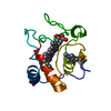
 Thermoplasma acidophilum DSM 1728 (acidophilic)
 X-RAY DIFFRACTION /
  Authors
 Citation
 Structure visualization
 Downloads & links
 Other downloads

 PDBj
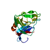
PDBj Assembly
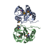


 Mass: 18.015 Da / Num. of mol.: 336 / Source method: isolated from a natural source / Formula: H2O
 Sample preparation
 / Beamline: 23-ID-D / Wavelength: 0.97945 Å
 Processing