 Movie
Movie Controller

[English] 日本語
 Yorodumi
- PDB-2r69: Crystal structure of Fab 1A1D-2 complexed with E-DIII of Dengue v... -
+ Open data

- Basic information
| Entry | Database: PDB / ID: 2r69 | ||||||
|---|---|---|---|---|---|---|---|
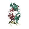
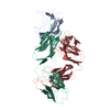
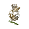
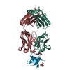
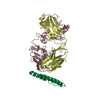
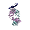
| Title | Crystal structure of Fab 1A1D-2 complexed with E-DIII of Dengue virus at 3.8 angstrom resolution | ||||||
 Components Components |
| ||||||
 Keywords Keywords | viral protein/Immune system / Fab-antigen complex / Capsid protein / Cleavage on pair of basic residues / Core protein / Envelope protein / Glycoprotein / Membrane / Transmembrane / Virion / viral protein-Immune system COMPLEX | ||||||
| Function / homology |  Function and homology information Function and homology informationsymbiont-mediated suppression of host interferon-mediated signaling pathway / flavivirin / symbiont-mediated suppression of host JAK-STAT cascade via inhibition of host TYK2 activity / host cell mitochondrion / symbiont-mediated suppression of host JAK-STAT cascade via inhibition of STAT2 activity / symbiont-mediated suppression of host cytoplasmic pattern recognition receptor signaling pathway via inhibition of MAVS activity / viral capsid / nucleoside-triphosphate phosphatase / double-stranded RNA binding / viral nucleocapsid ...symbiont-mediated suppression of host interferon-mediated signaling pathway / flavivirin / symbiont-mediated suppression of host JAK-STAT cascade via inhibition of host TYK2 activity / host cell mitochondrion / symbiont-mediated suppression of host JAK-STAT cascade via inhibition of STAT2 activity / symbiont-mediated suppression of host cytoplasmic pattern recognition receptor signaling pathway via inhibition of MAVS activity / viral capsid / nucleoside-triphosphate phosphatase / double-stranded RNA binding / viral nucleocapsid / channel activity / monoatomic ion transmembrane transport / clathrin-dependent endocytosis of virus by host cell / mRNA (guanine-N7)-methyltransferase / methyltransferase cap1 / host cell cytoplasm / methyltransferase cap1 activity / protein-macromolecule adaptor activity / mRNA 5'-cap (guanine-N7-)-methyltransferase activity / RNA helicase activity / protein dimerization activity / symbiont-mediated suppression of host innate immune response / host cell perinuclear region of cytoplasm / host cell endoplasmic reticulum membrane / RNA helicase / symbiont-mediated suppression of host type I interferon-mediated signaling pathway / serine-type endopeptidase activity / symbiont-mediated activation of host autophagy / RNA-directed RNA polymerase / viral RNA genome replication / RNA-directed RNA polymerase activity / fusion of virus membrane with host endosome membrane / viral envelope / lipid binding / virion attachment to host cell / host cell nucleus / virion membrane / structural molecule activity / ATP hydrolysis activity / proteolysis / extracellular region / ATP binding / metal ion binding Similarity search - Function | ||||||
| Biological species |  Dengue virus 2 Thailand/16681/84 Dengue virus 2 Thailand/16681/84 | ||||||
| Method |  X-RAY DIFFRACTION / SYNCHROTRON / MOLECULAR REPLACEMENT / molecular replacement / Resolution: 3.8 Å X-RAY DIFFRACTION / SYNCHROTRON / MOLECULAR REPLACEMENT / molecular replacement / Resolution: 3.8 Å | ||||||
 Authors Authors | Lok, S.M. / Kostyuchenko, V.K. / Nybakken, G.E. / Holdaway, H.A. / Battisti, A.J. / Sukupolvi-petty, S. / Sedlak, D. / Fremont, D.H. / Chipman, P.R. / Roehrig, J.T. ...Lok, S.M. / Kostyuchenko, V.K. / Nybakken, G.E. / Holdaway, H.A. / Battisti, A.J. / Sukupolvi-petty, S. / Sedlak, D. / Fremont, D.H. / Chipman, P.R. / Roehrig, J.T. / Diamond, M.S. / Kuhn, R.J. / Rossmann, M.G. | ||||||
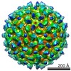
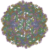
 Citation Citation | Journal: Nat Struct Mol Biol / Year: 2008 Title: Binding of a neutralizing antibody to dengue virus alters the arrangement of surface glycoproteins. Authors: Shee-Mei Lok / Victor Kostyuchenko / Grant E Nybakken / Heather A Holdaway / Anthony J Battisti / Soila Sukupolvi-Petty / Dagmar Sedlak / Daved H Fremont / Paul R Chipman / John T Roehrig / ...Authors: Shee-Mei Lok / Victor Kostyuchenko / Grant E Nybakken / Heather A Holdaway / Anthony J Battisti / Soila Sukupolvi-Petty / Dagmar Sedlak / Daved H Fremont / Paul R Chipman / John T Roehrig / Michael S Diamond / Richard J Kuhn / Michael G Rossmann /  Abstract: The monoclonal antibody 1A1D-2 has been shown to strongly neutralize dengue virus serotypes 1, 2 and 3, primarily by inhibiting attachment to host cells. A crystal structure of its antigen binding ...The monoclonal antibody 1A1D-2 has been shown to strongly neutralize dengue virus serotypes 1, 2 and 3, primarily by inhibiting attachment to host cells. A crystal structure of its antigen binding fragment (Fab) complexed with domain III of the viral envelope glycoprotein, E, showed that the epitope would be partially occluded in the known structure of the mature dengue virus. Nevertheless, antibody could bind to the virus at 37 degrees C, suggesting that the virus is in dynamic motion making hidden epitopes briefly available. A cryo-electron microscope image reconstruction of the virus:Fab complex showed large changes in the organization of the E protein that exposed the epitopes on two of the three E molecules in each of the 60 icosahedral asymmetric units of the virus. The changes in the structure of the viral surface are presumably responsible for inhibiting attachment to cells. | ||||||
| History |
|
- Structure visualization
| Structure viewer | Molecule: MolmilJmol/JSmol |
|---|
- Downloads & links
-Download
| PDBx/mmCIF format | 2r69.cif.gz | 98.5 KB | Display | PDBx/mmCIF format |
|---|---|---|---|---|
| PDB format | pdb2r69.ent.gz | 72.1 KB | Display | PDB format |
| PDBx/mmJSON format | 2r69.json.gz | Tree view | PDBx/mmJSON format | |
| Others |  Other downloads Other downloads |
-Validation report
| Arichive directory | https://data.pdbj.org/pub/pdb/validation_reports/r6/2r69ftp://data.pdbj.org/pub/pdb/validation_reports/r6/2r69 | HTTPS FTP |
|---|
-Related structure data


-Links
 PDBj
PDBj


- Assembly
| Deposited unit | 
| ||||||||
|---|---|---|---|---|---|---|---|---|---|
| 1 |
| ||||||||
| Unit cell |
|
-Components
| #1: Protein | Mass: 10932.665 Da / Num. of mol.: 1 Source method: isolated from a genetically manipulated source Source: (gene. exp.) Dengue virus 2 Thailand/16681/84 / Genus: Flavivirus / Species: Dengue virus / Strain: 16681 / Gene: E protein / Plasmid: pET21 / Species (production host): Escherichia coli / Production host:  |
|---|---|
| #2: Antibody | Mass: 22926.557 Da / Num. of mol.: 1 / Fragment: Fab Source method: isolated from a genetically manipulated source Source: (gene. exp.) |
| #3: Antibody | Mass: 23412.869 Da / Num. of mol.: 1 / Fragment: Fab Source method: isolated from a genetically manipulated source Source: (gene. exp.) |
| Has protein modification | Y |
-Experimental details
-Experiment
| Experiment | Method: X-RAY DIFFRACTION / Number of used crystals: 1 |
|---|
- Sample preparation
| Crystal | Density Matthews: 3.52 Å3/Da / Density % sol: 65.01 % |
|---|---|
| Crystal grow | Temperature: 298 K / Method: vapor diffusion, hanging drop / pH: 5.8 Details: 12% PEG 3350, pH 5.8, VAPOR DIFFUSION, HANGING DROP, temperature 298K |
-Data collection
| Diffraction | Mean temperature: 100 K |
|---|---|
| Diffraction source | Source: SYNCHROTRON / Site: APS / Beamline: 23-ID-D / Wavelength: 0.97857 Å |
| Detector | Type: MARMOSAIC 300 mm CCD / Detector: CCD / Date: Jan 1, 2005 |
| Radiation | Protocol: SINGLE WAVELENGTH / Monochromatic (M) / Laue (L): M / Scattering type: x-ray |
| Radiation wavelength | Wavelength: 0.97857 Å / Relative weight: 1 |
| Reflection | Resolution: 3.6→50 Å / Num. obs: 6433 / % possible obs: 68.3 % / Redundancy: 2.3 % / Rmerge(I) obs: 0.118 / Χ2: 1.226 / Net I/σ(I): 8 |
| Reflection shell | Resolution: 3.6→3.73 Å / Redundancy: 1.6 % / Rmerge(I) obs: 0.219 / Num. unique all: 352 / Χ2: 1.123 / % possible all: 37.4 |
-Phasing
| Phasing | Method: molecular replacement | |||||||||
|---|---|---|---|---|---|---|---|---|---|---|
| Phasing MR | Rfactor: 0.458 / Cor.coef. Fo:Fc: 0.31
|
- Processing
| Software |
| ||||||||||||||||||||||||||||
|---|---|---|---|---|---|---|---|---|---|---|---|---|---|---|---|---|---|---|---|---|---|---|---|---|---|---|---|---|---|
| Refinement | Method to determine structure: MOLECULAR REPLACEMENT / Resolution: 3.8→20 Å / σ(F): 0
| ||||||||||||||||||||||||||||
| Solvent computation | Bsol: 10 Å2 | ||||||||||||||||||||||||||||
| Displacement parameters | Biso mean: 19.789 Å2
| ||||||||||||||||||||||||||||
| Refinement step | Cycle: LAST / Resolution: 3.8→20 Å
| ||||||||||||||||||||||||||||
| Xplor file | Serial no: 1 / Param file: CNS_TOPPAR:protein_rep.param |
