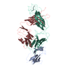
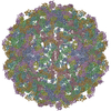
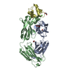
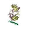
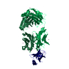
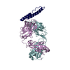
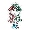
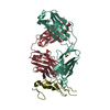
Journal: Nat Struct Mol Biol / Year: 2008 Title: Binding of a neutralizing antibody to dengue virus alters the arrangement of surface glycoproteins. Authors: Shee-Mei Lok / Victor Kostyuchenko / Grant E Nybakken / Heather A Holdaway / Anthony J Battisti / Soila Sukupolvi-Petty / Dagmar Sedlak / Daved H Fremont / Paul R Chipman / John T Roehrig / ...Authors: Shee-Mei Lok / Victor Kostyuchenko / Grant E Nybakken / Heather A Holdaway / Anthony J Battisti / Soila Sukupolvi-Petty / Dagmar Sedlak / Daved H Fremont / Paul R Chipman / John T Roehrig / Michael S Diamond / Richard J Kuhn / Michael G Rossmann / Abstract: The monoclonal antibody 1A1D-2 has been shown to strongly neutralize dengue virus serotypes 1, 2 and 3, primarily by inhibiting attachment to host cells. A crystal structure of its antigen binding ...The monoclonal antibody 1A1D-2 has been shown to strongly neutralize dengue virus serotypes 1, 2 and 3, primarily by inhibiting attachment to host cells. A crystal structure of its antigen binding fragment (Fab) complexed with domain III of the viral envelope glycoprotein, E, showed that the epitope would be partially occluded in the known structure of the mature dengue virus. Nevertheless, antibody could bind to the virus at 37 degrees C, suggesting that the virus is in dynamic motion making hidden epitopes briefly available. A cryo-electron microscope image reconstruction of the virus:Fab complex showed large changes in the organization of the E protein that exposed the epitopes on two of the three E molecules in each of the 60 icosahedral asymmetric units of the virus. The changes in the structure of the viral surface are presumably responsible for inhibiting attachment to cells.
In the structure databanks used in Yorodumi, some data are registered as the other names, "COVID-19 virus" and "2019-nCoV". Here are the details of the virus and the list of structure data.
Jan 31, 2019. EMDB accession codes are about to change! (news from PDBe EMDB page)
EMDB accession codes are about to change! (news from PDBe EMDB page)
The allocation of 4 digits for EMDB accession codes will soon come to an end. Whilst these codes will remain in use, new EMDB accession codes will include an additional digit and will expand incrementally as the available range of codes is exhausted. The current 4-digit format prefixed with “EMD-” (i.e. EMD-XXXX) will advance to a 5-digit format (i.e. EMD-XXXXX), and so on. It is currently estimated that the 4-digit codes will be depleted around Spring 2019, at which point the 5-digit format will come into force.
The EM Navigator/Yorodumi systems omit the EMD- prefix.
Related info.:Q: What is EMD? / ID/Accession-code notation in Yorodumi/EM Navigator
Yorodumi is a browser for structure data from EMDB, PDB, SASBDB, etc.
This page is also the successor to EM Navigator detail page, and also detail information page/front-end page for Omokage search.
The word "yorodu" (or yorozu) is an old Japanese word meaning "ten thousand". "mi" (miru) is to see.
Related info.:EMDB / PDB / SASBDB / Comparison of 3 databanks / Yorodumi Search / Aug 31, 2016. New EM Navigator & Yorodumi / Yorodumi Papers / Jmol/JSmol / Function and homology information / Changes in new EM Navigator and Yorodumi
 Movie
Movie Controller

 Yorodumi
 Open data
 Basic information
 Components
 Keywords
 Function and homology information
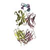
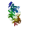
 Dengue virus 2 Thailand/16681/84
 X-RAY DIFFRACTION /
  Authors
 Citation
 Structure visualization
 Downloads & links
 Other downloads
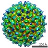


 PDBj
PDBj


 Assembly
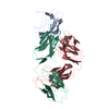

 Mass: 18.015 Da / Num. of mol.: 254 / Source method: isolated from a natural source / Formula: H2O
 Sample preparation
 Processing