 Movie
Movie Controller

[English] 日本語
 Yorodumi
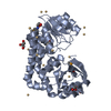
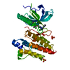
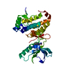
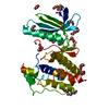
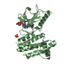
- PDB-2qon: Human EphA3 kinase and juxtamembrane region, Y596F:Y602F:Y742A tr... -
+ Open data

- Basic information
| Entry | Database: PDB / ID: 2qon | ||||||
|---|---|---|---|---|---|---|---|
| Title | Human EphA3 kinase and juxtamembrane region, Y596F:Y602F:Y742A triple mutant | ||||||
 Components Components | Ephrin receptor | ||||||
 Keywords Keywords | TRANSFERASE / receptor tyrosine kinase / juxtamembrane segment / structural genomics / mutant / Structural Genomics Consortium / SGC / ATP-binding / Nucleotide-binding / Phosphorylation / Transmembrane / Tyrosine-protein kinase | ||||||
| Function / homology |  Function and homology information Function and homology informationfasciculation of motor neuron axon / fasciculation of sensory neuron axon / transmembrane-ephrin receptor activity / GPI-linked ephrin receptor activity / regulation of epithelial to mesenchymal transition / ephrin receptor activity / negative regulation of endocytosis / EPH-Ephrin signaling / regulation of focal adhesion assembly / negative regulation of epithelial to mesenchymal transition ...fasciculation of motor neuron axon / fasciculation of sensory neuron axon / transmembrane-ephrin receptor activity / GPI-linked ephrin receptor activity / regulation of epithelial to mesenchymal transition / ephrin receptor activity / negative regulation of endocytosis / EPH-Ephrin signaling / regulation of focal adhesion assembly / negative regulation of epithelial to mesenchymal transition / EPHA-mediated growth cone collapse / regulation of cell-cell adhesion / EPH-ephrin mediated repulsion of cells / ephrin receptor signaling pathway / cellular response to retinoic acid / peptidyl-tyrosine phosphorylation / axon guidance / regulation of actin cytoskeleton organization / regulation of microtubule cytoskeleton organization / positive regulation of protein localization to plasma membrane / positive regulation of neuron projection development / receptor protein-tyrosine kinase / cell migration / early endosome / cell adhesion / dendrite / extracellular region / ATP binding / plasma membrane Similarity search - Function | ||||||
| Biological species |  Homo sapiens (human) Homo sapiens (human) | ||||||
| Method |  X-RAY DIFFRACTION / SYNCHROTRON / MOLECULAR REPLACEMENT / molecular replacement / Resolution: 1.79 Å X-RAY DIFFRACTION / SYNCHROTRON / MOLECULAR REPLACEMENT / molecular replacement / Resolution: 1.79 Å | ||||||
 Authors Authors | Davis, T. / Walker, J.R. / Newman, E.M. / Mackenzie, F. / Butler-Cole, C. / Weigelt, J. / Sundstrom, M. / Arrowsmith, C.H. / Edwards, A.M. / Bochkarev, A. ...Davis, T. / Walker, J.R. / Newman, E.M. / Mackenzie, F. / Butler-Cole, C. / Weigelt, J. / Sundstrom, M. / Arrowsmith, C.H. / Edwards, A.M. / Bochkarev, A. / Dhe-Paganon, S. / Structural Genomics Consortium (SGC) | ||||||
 Citation Citation | Journal: Structure / Year: 2008 Title: Autoregulation by the Juxtamembrane Region of the Human Ephrin Receptor Tyrosine Kinase A3 (EphA3). Authors: Davis, T.L. / Walker, J.R. / Loppnau, P. / Butler-Cole, C. / Allali-Hassani, A. / Dhe-Paganon, S. | ||||||
| History |
|
- Structure visualization
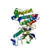
| Structure viewer | Molecule: MolmilJmol/JSmol |
|---|
- Downloads & links
-Download
| PDBx/mmCIF format | 2qon.cif.gz | 81.6 KB | Display | PDBx/mmCIF format |
|---|---|---|---|---|
| PDB format | pdb2qon.ent.gz | 59.3 KB | Display | PDB format |
| PDBx/mmJSON format | 2qon.json.gz | Tree view | PDBx/mmJSON format | |
| Others |  Other downloads Other downloads |
-Validation report
| Arichive directory | https://data.pdbj.org/pub/pdb/validation_reports/qo/2qonftp://data.pdbj.org/pub/pdb/validation_reports/qo/2qon | HTTPS FTP |
|---|
-Related structure data
| Related structure data |  2qo2C  2qo7C  2qo9C  2qobC  2qocC  2qodC  2qofC  2qoiC  2qokC  2qolC  2qooC  2qoqC  2gsfS S: Starting model for refinement C: citing same article ( |
|---|---|
| Similar structure data |


-Links
 PDBj
PDBj


- Assembly
| Deposited unit | 
| ||||||||
|---|---|---|---|---|---|---|---|---|---|
| 1 |
| ||||||||
| Unit cell |
|
-Components
| #1: Protein | Mass: 41919.746 Da / Num. of mol.: 1 Fragment: Juxtamembrane segment and kinase domain: Residues 577-947 Mutation: Y596F, Y602F, Y742A Source method: isolated from a genetically manipulated source Source: (gene. exp.) Homo sapiens (human) / Tissue: Placenta / Gene: EPHA3 / Plasmid: pET28a-LIC / Species (production host): Escherichia coli / Production host:  References: UniProt: Q6P4R6, UniProt: P29320*PLUS, receptor protein-tyrosine kinase |
|---|---|
| #2: Chemical | ChemComp-GOL /   Mass: 92.094 Da / Num. of mol.: 1 / Source method: obtained synthetically / Formula: C3H8O3 Mass: 92.094 Da / Num. of mol.: 1 / Source method: obtained synthetically / Formula: C3H8O3 |
| #3: Water | ChemComp-HOH /  Mass: 18.015 Da / Num. of mol.: 333 / Source method: isolated from a natural source / Formula: H2O Mass: 18.015 Da / Num. of mol.: 333 / Source method: isolated from a natural source / Formula: H2O |
| Has protein modification | Y |
-Experimental details
-Experiment
| Experiment | Method: X-RAY DIFFRACTION / Number of used crystals: 1 |
|---|
- Sample preparation
| Crystal | Density Matthews: 1.86 Å3/Da / Density % sol: 33.81 % |
|---|---|
| Crystal grow | Temperature: 298 K / Method: vapor diffusion, sitting drop / pH: 7.5 Details: 20 mg/mL Protein, 25% PEG 3350, 0.2M Ammonium sulfate, 0.1M Hepes, pH 7.5, VAPOR DIFFUSION, SITTING DROP, temperature 298K |
-Data collection
| Diffraction | Mean temperature: 100 K | |||||||||||||||||||||||||||||||||||||||||||||||||||||||
|---|---|---|---|---|---|---|---|---|---|---|---|---|---|---|---|---|---|---|---|---|---|---|---|---|---|---|---|---|---|---|---|---|---|---|---|---|---|---|---|---|---|---|---|---|---|---|---|---|---|---|---|---|---|---|---|---|
| Diffraction source | Source: SYNCHROTRON / Site: APS  / Beamline: 17-ID / Wavelength: 1 Å / Beamline: 17-ID / Wavelength: 1 Å | |||||||||||||||||||||||||||||||||||||||||||||||||||||||
| Detector | Type: ADSC QUANTUM 210 / Detector: CCD / Date: Oct 22, 2006 | |||||||||||||||||||||||||||||||||||||||||||||||||||||||
| Radiation | Protocol: SINGLE WAVELENGTH / Monochromatic (M) / Laue (L): M / Scattering type: x-ray | |||||||||||||||||||||||||||||||||||||||||||||||||||||||
| Radiation wavelength | Wavelength: 1 Å / Relative weight: 1 | |||||||||||||||||||||||||||||||||||||||||||||||||||||||
| Reflection | Resolution: 1.79→40 Å / Num. obs: 26894 / % possible obs: 92 % / Redundancy: 4.1 % / Rmerge(I) obs: 0.057 / Net I/σ(I): 11.8 | |||||||||||||||||||||||||||||||||||||||||||||||||||||||
| Reflection shell |
|
-Phasing
| Phasing | Method: molecular replacement |
|---|
- Processing
| Software |
| ||||||||||||||||||||||||||||||||||||||||||||||||||||||||||||||||||||||||||||||||||||||||||||||||||||||||||||||||||||||||||||||||||||||||||||||||||||||||||||||||||||||||||
|---|---|---|---|---|---|---|---|---|---|---|---|---|---|---|---|---|---|---|---|---|---|---|---|---|---|---|---|---|---|---|---|---|---|---|---|---|---|---|---|---|---|---|---|---|---|---|---|---|---|---|---|---|---|---|---|---|---|---|---|---|---|---|---|---|---|---|---|---|---|---|---|---|---|---|---|---|---|---|---|---|---|---|---|---|---|---|---|---|---|---|---|---|---|---|---|---|---|---|---|---|---|---|---|---|---|---|---|---|---|---|---|---|---|---|---|---|---|---|---|---|---|---|---|---|---|---|---|---|---|---|---|---|---|---|---|---|---|---|---|---|---|---|---|---|---|---|---|---|---|---|---|---|---|---|---|---|---|---|---|---|---|---|---|---|---|---|---|---|---|---|---|
| Refinement | Method to determine structure: MOLECULAR REPLACEMENT Starting model: PDB entry 2GSF Resolution: 1.79→34.18 Å / Cor.coef. Fo:Fc: 0.957 / Cor.coef. Fo:Fc free: 0.949 / SU B: 2.486 / SU ML: 0.077 / Cross valid method: THROUGHOUT / σ(F): 0 / ESU R: 0.137 / ESU R Free: 0.115 Stereochemistry target values: MAXIMUM LIKELIHOOD WITH PHASES Details: HYDROGENS HAVE BEEN ADDED IN THE RIDING POSITIONS
| ||||||||||||||||||||||||||||||||||||||||||||||||||||||||||||||||||||||||||||||||||||||||||||||||||||||||||||||||||||||||||||||||||||||||||||||||||||||||||||||||||||||||||
| Solvent computation | Ion probe radii: 0.8 Å / Shrinkage radii: 0.8 Å / VDW probe radii: 1.2 Å / Solvent model: MASK | ||||||||||||||||||||||||||||||||||||||||||||||||||||||||||||||||||||||||||||||||||||||||||||||||||||||||||||||||||||||||||||||||||||||||||||||||||||||||||||||||||||||||||
| Displacement parameters | Biso mean: 17.312 Å2
| ||||||||||||||||||||||||||||||||||||||||||||||||||||||||||||||||||||||||||||||||||||||||||||||||||||||||||||||||||||||||||||||||||||||||||||||||||||||||||||||||||||||||||
| Refinement step | Cycle: LAST / Resolution: 1.79→34.18 Å
| ||||||||||||||||||||||||||||||||||||||||||||||||||||||||||||||||||||||||||||||||||||||||||||||||||||||||||||||||||||||||||||||||||||||||||||||||||||||||||||||||||||||||||
| Refine LS restraints |
| ||||||||||||||||||||||||||||||||||||||||||||||||||||||||||||||||||||||||||||||||||||||||||||||||||||||||||||||||||||||||||||||||||||||||||||||||||||||||||||||||||||||||||
| LS refinement shell | Resolution: 1.79→1.84 Å / Total num. of bins used: 20
|
