- PDB-2qjz: Structural Basis of Microtubule Plus End Tracking by XMAP215, CLI... -
+
Open data
ID or keywords:
Loading...
-
Basic information
Entry
Database: PDB / ID: 2qjz
Title
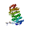
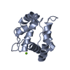
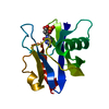
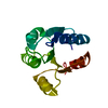
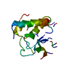
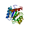
Structural Basis of Microtubule Plus End Tracking by XMAP215, CLIP-170 and EB1
Components
Microtubule-associated protein RP/EB family member 1
Keywords
PROTEIN BINDING / EB1 / calponin homology domain / microtubule plus end / +TIP
Function / homology
Function and homology information
protein localization to astral microtubule / protein localization to mitotic spindle / cortical microtubule cytoskeleton / mitotic spindle astral microtubule end / protein localization to microtubule / cell projection membrane / microtubule plus-end / mitotic spindle microtubule / attachment of mitotic spindle microtubules to kinetochore / microtubule plus-end binding ...protein localization to astral microtubule / protein localization to mitotic spindle / cortical microtubule cytoskeleton / mitotic spindle astral microtubule end / protein localization to microtubule / cell projection membrane / microtubule plus-end / mitotic spindle microtubule / attachment of mitotic spindle microtubules to kinetochore / microtubule plus-end binding / microtubule bundle formation / non-motile cilium assembly / protein localization to centrosome / mitotic spindle pole / spindle midzone / establishment of mitotic spindle orientation / negative regulation of microtubule polymerization / microtubule polymerization / microtubule organizing center / regulation of microtubule polymerization or depolymerization / positive regulation of microtubule polymerization / cytoplasmic microtubule / spindle assembly / Loss of Nlp from mitotic centrosomes / Loss of proteins required for interphase microtubule organization from the centrosome / Amplification of signal from unattached kinetochores via a MAD2 inhibitory signal / Recruitment of mitotic centrosome proteins and complexes / Recruitment of NuMA to mitotic centrosomes / Anchoring of the basal body to the plasma membrane / Mitotic Prometaphase / EML4 and NUDC in mitotic spindle formation / AURKA Activation by TPX2 / Resolution of Sister Chromatid Cohesion / protein serine/threonine kinase binding / RHO GTPases Activate Formins / intracellular protein localization / The role of GTSE1 in G2/M progression after G2 checkpoint / Separation of Sister Chromatids / Regulation of PLK1 Activity at G2/M Transition / cell migration / microtubule / ciliary basal body / cadherin binding / cell division / focal adhesion / centrosome / Golgi apparatus / RNA binding / identical protein binding / cytosol Similarity search - Function
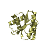
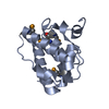
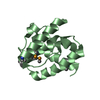
The biological assembly is a dimer formed by a conserved C-terminal coiled-coil / four helix bundle dimerization domain not included in this construct.
-
Components
#1: Protein
Microtubule-associatedproteinRP/EBfamilymember1 / APC-binding protein EB1 / End-binding protein 1 / EB1
Mass: 14455.015 Da / Num. of mol.: 2 / Fragment: Calponin Homology Domain Source method: isolated from a genetically manipulated source Source: (gene. exp.) Homo sapiens (human) / Gene: MAPRE1 / Plasmid: pGEX-6P2 / Production host: Escherichia coli (E. coli) / Strain (production host): BL21 DE3 pLysS / References: UniProt: Q15691
In the structure databanks used in Yorodumi, some data are registered as the other names, "COVID-19 virus" and "2019-nCoV". Here are the details of the virus and the list of structure data.
Jan 31, 2019. EMDB accession codes are about to change! (news from PDBe EMDB page)
EMDB accession codes are about to change! (news from PDBe EMDB page)
The allocation of 4 digits for EMDB accession codes will soon come to an end. Whilst these codes will remain in use, new EMDB accession codes will include an additional digit and will expand incrementally as the available range of codes is exhausted. The current 4-digit format prefixed with “EMD-” (i.e. EMD-XXXX) will advance to a 5-digit format (i.e. EMD-XXXXX), and so on. It is currently estimated that the 4-digit codes will be depleted around Spring 2019, at which point the 5-digit format will come into force.
The EM Navigator/Yorodumi systems omit the EMD- prefix.
Related info.:Q: What is EMD? / ID/Accession-code notation in Yorodumi/EM Navigator
Yorodumi is a browser for structure data from EMDB, PDB, SASBDB, etc.
This page is also the successor to EM Navigator detail page, and also detail information page/front-end page for Omokage search.
The word "yorodu" (or yorozu) is an old Japanese word meaning "ten thousand". "mi" (miru) is to see.
Related info.:EMDB / PDB / SASBDB / Comparison of 3 databanks / Yorodumi Search / Aug 31, 2016. New EM Navigator & Yorodumi / Yorodumi Papers / Jmol/JSmol / Function and homology information / Changes in new EM Navigator and Yorodumi
 Movie
Movie Controller

 Yorodumi
 Open data
 Basic information
 Components
 Keywords
 Function and homology information
 Homo sapiens (human)
 X-RAY DIFFRACTION /
  Authors
 Citation
 Structure visualization
 Downloads & links
 Other downloads
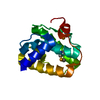

 PDBj
PDBj


 Assembly
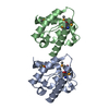

 Mass: 18.015 Da / Num. of mol.: 302 / Source method: isolated from a natural source / Formula: H2O
 Sample preparation
 / Beamline: 8.3.1 / Wavelength: 0.9798, 1.1271, 0.7999
 Processing