 Movie
Movie Controller


+ Open data

- Basic information
| Entry | Database: PDB / ID: 2p30 | ||||||
|---|---|---|---|---|---|---|---|
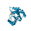
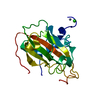
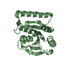
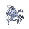
| Title | Crystal structure of TTHB049 from Thermus thermophilus HB8 | ||||||
 Components Components | Alpha-ribazole-5'-phosphate phosphatase | ||||||
 Keywords Keywords | HYDROLASE / Thermus thermophilus HB8 / Structural Genomics / NPPSFA / National Project on Protein Structural and Functional Analyses / RIKEN Structural Genomics/Proteomics Initiative / RSGI | ||||||
| Function / homology |  Function and homology information Function and homology informationphosphatase activity / isomerase activity / glycolytic process / cytoplasm Similarity search - Function | ||||||
| Biological species |   Thermus thermophilus (bacteria) Thermus thermophilus (bacteria) | ||||||
| Method |  X-RAY DIFFRACTION / FOURIER SYNTHESIS / Resolution: 1.85 Å X-RAY DIFFRACTION / FOURIER SYNTHESIS / Resolution: 1.85 Å | ||||||
 Authors Authors | Sugahara, M. / Taketa, M. / Morikawa, Y. / Matsuura, Y. / Kunishima, N. / RIKEN Structural Genomics/Proteomics Initiative (RSGI) | ||||||
 Citation Citation | Journal: To be Published Title: Crystal structure of TTHB049 from Thermus thermophilus HB8 Authors: Sugahara, M. / Taketa, M. / Morikawa, Y. / Matsuura, Y. / Kunishima, N. | ||||||
| History |
|
- Structure visualization
| Structure viewer | Molecule: MolmilJmol/JSmol |
|---|
- Downloads & links
-Download
| PDBx/mmCIF format | 2p30.cif.gz | 50.7 KB | Display | PDBx/mmCIF format |
|---|---|---|---|---|
| PDB format | pdb2p30.ent.gz | 35.6 KB | Display | PDB format |
| PDBx/mmJSON format | 2p30.json.gz | Tree view | PDBx/mmJSON format | |
| Others |  Other downloads Other downloads |
-Validation report
| Arichive directory | https://data.pdbj.org/pub/pdb/validation_reports/p3/2p30ftp://data.pdbj.org/pub/pdb/validation_reports/p3/2p30 | HTTPS FTP |
|---|
-Related structure data
| Related structure data | 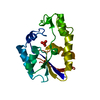 2hiaC 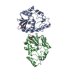 2owdC 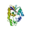 2oweC 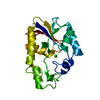 2p2yC 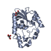 2p2zC 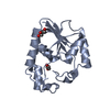 2p6mC 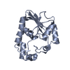 2p6oC 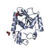 2p75C 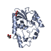 2p77C 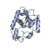 2p78C 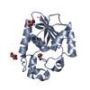 2p79C 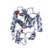 2p9fC 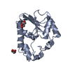 2p9yC 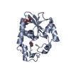 2p9zC 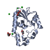 2pa0C 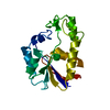 1v7qS C: citing same article ( S: Starting model for refinement |
|---|---|
| Similar structure data | |
| Other databases |


-Links
 PDBj
PDBj


- Assembly
| Deposited unit | 
| ||||||||
|---|---|---|---|---|---|---|---|---|---|
| 1 |
| ||||||||
| 2 | 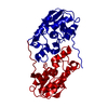
| ||||||||
| Unit cell |
| ||||||||
| Details | The biological assembly is a monomer in the asymmetric unit |
-Components
| #1: Protein | Mass: 19646.502 Da / Num. of mol.: 1 / Mutation: L170M Source method: isolated from a genetically manipulated source Source: (gene. exp.) Thermus thermophilus (bacteria) / Plasmid: pET-11A / Species (production host): Escherichia coli / Production host: References: UniProt: Q53WB3, adenosylcobalamin/alpha-ribazole phosphatase |
|---|---|
| #2: Water | ChemComp-HOH /  Mass: 18.015 Da / Num. of mol.: 160 / Source method: isolated from a natural source / Formula: H2O Mass: 18.015 Da / Num. of mol.: 160 / Source method: isolated from a natural source / Formula: H2O |
-Experimental details
-Experiment
| Experiment | Method: X-RAY DIFFRACTION / Number of used crystals: 1 |
|---|
- Sample preparation
| Crystal | Density Matthews: 1.81 Å3/Da / Density % sol: 32.22 % |
|---|---|
| Crystal grow | Temperature: 295 K / Method: oil micro batch / pH: 4.8 Details: 2.75M NaCl, 0.1M citrate, pH 4.8, oil micro batch, temperature 295K |
-Data collection
| Diffraction | Mean temperature: 100 K |
|---|---|
| Diffraction source | Source: ROTATING ANODE / Type: RIGAKU / Wavelength: 1.54 Å |
| Detector | Type: RIGAKU RAXIS IV / Detector: IMAGE PLATE / Date: Dec 25, 2006 |
| Radiation | Protocol: SINGLE WAVELENGTH / Monochromatic (M) / Laue (L): M / Scattering type: x-ray |
| Radiation wavelength | Wavelength: 1.54 Å / Relative weight: 1 |
| Reflection | Resolution: 1.85→20 Å / Num. all: 13233 / Num. obs: 13233 / % possible obs: 100 % / Observed criterion σ(F): 0 / Observed criterion σ(I): 0 / Redundancy: 12.3 % / Biso Wilson estimate: 22.3 Å2 / Rmerge(I) obs: 0.048 / Rsym value: 0.046 / Net I/σ(I): 13.9 |
| Reflection shell | Resolution: 1.85→1.92 Å / Redundancy: 11.5 % / Rmerge(I) obs: 0.199 / Mean I/σ(I) obs: 8 / Num. unique all: 1266 / Rsym value: 0.192 / % possible all: 100 |
- Processing
| Software |
| |||||||||||||||||||||||||
|---|---|---|---|---|---|---|---|---|---|---|---|---|---|---|---|---|---|---|---|---|---|---|---|---|---|---|
| Refinement | Method to determine structure: FOURIER SYNTHESIS Starting model: 1v7q Resolution: 1.85→19.84 Å / Isotropic thermal model: Anisotrop / Cross valid method: THROUGHOUT / σ(F): 0 / Stereochemistry target values: Engh & Huber
| |||||||||||||||||||||||||
| Displacement parameters | Biso mean: 23.3 Å2
| |||||||||||||||||||||||||
| Refine analyze |
| |||||||||||||||||||||||||
| Refinement step | Cycle: LAST / Resolution: 1.85→19.84 Å
| |||||||||||||||||||||||||
| Refine LS restraints |
| |||||||||||||||||||||||||
| LS refinement shell | Resolution: 1.85→1.92 Å / Rfactor Rfree error: 0.033
|
