 Movie
Movie Controller

[English] 日本語
 Yorodumi
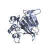
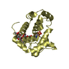
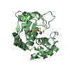
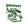
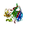
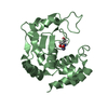
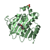
- PDB-4l5h: Structure of haze forming proteins in white wines: Vitis vinifera... -
+ Open data

- Basic information
| Entry | Database: PDB / ID: 4l5h | |||||||||
|---|---|---|---|---|---|---|---|---|---|---|
| Title | Structure of haze forming proteins in white wines: Vitis vinifera thaumatin-like proteins | |||||||||
 Components Components | VVTL1 | |||||||||
 Keywords Keywords | ANTIFUNGAL PROTEIN / PLANT PROTEIN | |||||||||
| Function / homology |  Function and homology information Function and homology informationThaumatin / Thaumatin / Thaumatin, conserved site / Thaumatin family signature. / Thaumatin family / Thaumatin family / Thaumatin family profile. / Thaumatin family / Osmotin/thaumatin-like superfamily / Sandwich / Mainly Beta Similarity search - Domain/homology | |||||||||
| Biological species |  | |||||||||
| Method |  X-RAY DIFFRACTION / SYNCHROTRON / MOLECULAR REPLACEMENT / Resolution: 1.8 Å X-RAY DIFFRACTION / SYNCHROTRON / MOLECULAR REPLACEMENT / Resolution: 1.8 Å | |||||||||
 Authors Authors | Marangon, M. / Menz, R.I. / Waters, E.J. / Van Sluyter, S.C. | |||||||||
 Citation Citation | Journal: Plos One / Year: 2014 Title: Structure of Haze Forming Proteins in White Wines: Vitis vinifera Thaumatin-Like Proteins. Authors: Marangon, M. / Van Sluyter, S.C. / Waters, E.J. / Menz, R.I. | |||||||||
| History |
|
- Structure visualization
| Structure viewer | Molecule: MolmilJmol/JSmol |
|---|
- Downloads & links
-Download
| PDBx/mmCIF format | 4l5h.cif.gz | 158.8 KB | Display | PDBx/mmCIF format |
|---|---|---|---|---|
| PDB format | pdb4l5h.ent.gz | 126.5 KB | Display | PDB format |
| PDBx/mmJSON format | 4l5h.json.gz | Tree view | PDBx/mmJSON format | |
| Others |  Other downloads Other downloads |
-Validation report
| Arichive directory | https://data.pdbj.org/pub/pdb/validation_reports/l5/4l5hftp://data.pdbj.org/pub/pdb/validation_reports/l5/4l5h | HTTPS FTP |
|---|
-Related structure data
| Related structure data |  4jruSC  4mbtC S: Starting model for refinement C: citing same article ( |
|---|---|
| Similar structure data |


-Links
 PDBj
PDBj
- Assembly
| Deposited unit | 
| ||||||||
|---|---|---|---|---|---|---|---|---|---|
| 1 | 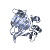
| ||||||||
| 2 | 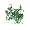
| ||||||||
| Unit cell |
|
-Components
| #1: Protein | Mass: 21301.609 Da / Num. of mol.: 2 / Fragment: UNP residues 25-222 / Source method: isolated from a natural source / Details: Grape juice / Source: (natural) #2: Chemical | ChemComp-GOL / |   Mass: 92.094 Da / Num. of mol.: 1 / Source method: obtained synthetically / Formula: C3H8O3 Mass: 92.094 Da / Num. of mol.: 1 / Source method: obtained synthetically / Formula: C3H8O3#3: Water | ChemComp-HOH / |  Mass: 18.015 Da / Num. of mol.: 181 / Source method: isolated from a natural source / Formula: H2O Mass: 18.015 Da / Num. of mol.: 181 / Source method: isolated from a natural source / Formula: H2OHas protein modification | Y | |
|---|
-Experimental details
-Experiment
| Experiment | Method: X-RAY DIFFRACTION / Number of used crystals: 1 |
|---|
- Sample preparation
| Crystal | Density Matthews: 2.64 Å3/Da / Density % sol: 53.46 % |
|---|---|
| Crystal grow | Temperature: 293 K / Method: vapor diffusion, sitting drop / pH: 4.6 Details: 0.1 M Na Acetate pH 4.6, 2 M Mg acetate, VAPOR DIFFUSION, SITTING DROP, temperature 293K |
-Data collection
| Diffraction | Mean temperature: 100 K | ||||||||||||||||||||||||||||||||||||||||||||||||||||||||||||||||||
|---|---|---|---|---|---|---|---|---|---|---|---|---|---|---|---|---|---|---|---|---|---|---|---|---|---|---|---|---|---|---|---|---|---|---|---|---|---|---|---|---|---|---|---|---|---|---|---|---|---|---|---|---|---|---|---|---|---|---|---|---|---|---|---|---|---|---|---|
| Diffraction source | Source: SYNCHROTRON / Site: Australian Synchrotron  / Beamline: MX1 / Wavelength: 0.956639 Å / Beamline: MX1 / Wavelength: 0.956639 Å | ||||||||||||||||||||||||||||||||||||||||||||||||||||||||||||||||||
| Detector | Type: ADSC QUANTUM 210r / Detector: CCD / Date: Nov 22, 2008 | ||||||||||||||||||||||||||||||||||||||||||||||||||||||||||||||||||
| Radiation | Protocol: SINGLE WAVELENGTH / Monochromatic (M) / Laue (L): M / Scattering type: x-ray | ||||||||||||||||||||||||||||||||||||||||||||||||||||||||||||||||||
| Radiation wavelength | Wavelength: 0.956639 Å / Relative weight: 1 | ||||||||||||||||||||||||||||||||||||||||||||||||||||||||||||||||||
| Reflection | Resolution: 1.59→12.63 Å / Num. obs: 29314 / % possible obs: 75.7 % / Observed criterion σ(I): 3 / Rmerge(I) obs: 0.081 / Net I/σ(I): 8.5 | ||||||||||||||||||||||||||||||||||||||||||||||||||||||||||||||||||
| Reflection shell |
|
- Processing
| Software |
| |||||||||||||||||||||||||||||||||||||||||||||
|---|---|---|---|---|---|---|---|---|---|---|---|---|---|---|---|---|---|---|---|---|---|---|---|---|---|---|---|---|---|---|---|---|---|---|---|---|---|---|---|---|---|---|---|---|---|---|
| Refinement | Method to determine structure: MOLECULAR REPLACEMENT Starting model: PDB ENTRY 4JRU Resolution: 1.8→12.63 Å / Cor.coef. Fo:Fc: 0.926 / Cor.coef. Fo:Fc free: 0.898 / SU B: 5.176 / SU ML: 0.079 / Cross valid method: THROUGHOUT / ESU R: 0.178 / ESU R Free: 0.156 / Stereochemistry target values: MAXIMUM LIKELIHOOD / Details: HYDROGENS HAVE BEEN USED IF PRESENT IN THE INPUT
| |||||||||||||||||||||||||||||||||||||||||||||
| Solvent computation | Ion probe radii: 0.8 Å / Shrinkage radii: 0.8 Å / VDW probe radii: 1.2 Å / Solvent model: MASK | |||||||||||||||||||||||||||||||||||||||||||||
| Displacement parameters | Biso mean: 12.485 Å2
| |||||||||||||||||||||||||||||||||||||||||||||
| Refinement step | Cycle: LAST / Resolution: 1.8→12.63 Å
| |||||||||||||||||||||||||||||||||||||||||||||
| Refine LS restraints |
| |||||||||||||||||||||||||||||||||||||||||||||
| LS refinement shell | Resolution: 1.8→1.846 Å / Total num. of bins used: 20
| |||||||||||||||||||||||||||||||||||||||||||||
| Refinement TLS params. | Method: refined / Origin x: -28.6775 Å / Origin y: 0.7956 Å / Origin z: -10.2748 Å
|
