 Movie
Movie Controller

[English] 日本語
 Yorodumi
- PDB-2od9: Structural Basis for Nicotinamide Inhibition and Base Exchange in... -
+ Open data

- Basic information
| Entry | Database: PDB / ID: 2od9 | ||||||
|---|---|---|---|---|---|---|---|
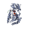
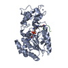
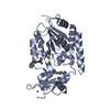
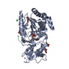
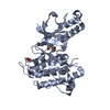
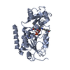
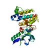
| Title | Structural Basis for Nicotinamide Inhibition and Base Exchange in Sir2 Enzymes | ||||||
 Components Components |
| ||||||
 Keywords Keywords | HYDROLASE / Zn binding protein / Rossmann fold | ||||||
| Function / homology |  Function and homology information Function and homology information: / negative regulation of mitotic recombination / histone H4K16 deacetylase activity, NAD-dependent / protein acetyllysine N-acetyltransferase / histone deacetylase activity, NAD-dependent / rDNA heterochromatin formation / NAD+ binding / metal ion binding / nucleus / cytoplasm Similarity search - Function | ||||||
| Biological species |  | ||||||
| Method |  X-RAY DIFFRACTION / SYNCHROTRON / MOLECULAR REPLACEMENT / Resolution: 2.05 Å X-RAY DIFFRACTION / SYNCHROTRON / MOLECULAR REPLACEMENT / Resolution: 2.05 Å | ||||||
 Authors Authors | Marmorstein, R. / Sanders, B.D. | ||||||
 Citation Citation | Journal: Mol.Cell / Year: 2007 Title: Structural basis for nicotinamide inhibition and base exchange in sir2 enzymes. Authors: Sanders, B.D. / Zhao, K. / Slama, J.T. / Marmorstein, R. | ||||||
| History |
|
- Structure visualization
| Structure viewer | Molecule: MolmilJmol/JSmol |
|---|
- Downloads & links
-Download
| PDBx/mmCIF format | 2od9.cif.gz | 80.3 KB | Display | PDBx/mmCIF format |
|---|---|---|---|---|
| PDB format | pdb2od9.ent.gz | 57.6 KB | Display | PDB format |
| PDBx/mmJSON format | 2od9.json.gz | Tree view | PDBx/mmJSON format | |
| Others |  Other downloads Other downloads |
-Validation report
| Arichive directory | https://data.pdbj.org/pub/pdb/validation_reports/od/2od9ftp://data.pdbj.org/pub/pdb/validation_reports/od/2od9 | HTTPS FTP |
|---|
-Related structure data
| Related structure data |  2od2C  2od7C  2qqfC  2qqgC 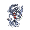 1szdS S: Starting model for refinement C: citing same article ( |
|---|---|
| Similar structure data |


-Links
 PDBj
PDBj

- Assembly
| Deposited unit | 
| ||||||||
|---|---|---|---|---|---|---|---|---|---|
| 1 |
| ||||||||
| 2 | 
| ||||||||
| Unit cell |
| ||||||||
| Details | The biological assembly is a trimer generated from the monomer in the asymmetric unit. |
-Components
-Protein / Protein/peptide , 2 types, 2 molecules AB
| #1: Protein | Mass: 34794.828 Da / Num. of mol.: 1 / Fragment: Hst2 catalytic core domain, residues 1-294 Source method: isolated from a genetically manipulated source Source: (gene. exp.) Gene: HST2 / Plasmid: pRSET / Production host:  References: UniProt: P53686, Hydrolases; Acting on carbon-nitrogen bonds, other than peptide bonds; In linear amides |
|---|---|
| #2: Protein/peptide | Mass: 1610.925 Da / Num. of mol.: 1 / Source method: obtained synthetically |
-Non-polymers , 4 types, 119 molecules 

| #3: Chemical | ChemComp-ZN /  Mass: 65.409 Da / Num. of mol.: 1 / Source method: obtained synthetically / Formula: Zn Mass: 65.409 Da / Num. of mol.: 1 / Source method: obtained synthetically / Formula: Zn |
|---|---|
| #4: Chemical | ChemComp-A1R /  Mass: 542.332 Da / Num. of mol.: 1 / Source method: obtained synthetically / Formula: C15H24N6O12P2 Mass: 542.332 Da / Num. of mol.: 1 / Source method: obtained synthetically / Formula: C15H24N6O12P2 |
| #5: Chemical | ChemComp-NCA /  Mass: 122.125 Da / Num. of mol.: 1 / Source method: obtained synthetically / Formula: C6H6N2O / Comment: medication*YM Mass: 122.125 Da / Num. of mol.: 1 / Source method: obtained synthetically / Formula: C6H6N2O / Comment: medication*YM |
| #6: Water | ChemComp-HOH / Mass: 18.015 Da / Num. of mol.: 116 / Source method: isolated from a natural source / Formula: H2O |
-Details
| Has protein modification | Y |
|---|
-Experimental details
-Experiment
| Experiment | Method: X-RAY DIFFRACTION / Number of used crystals: 1 |
|---|
- Sample preparation
| Crystal | Density Matthews: 3.12 Å3/Da / Density % sol: 60.61 % |
|---|---|
| Crystal grow | Temperature: 298 K / Method: vapor diffusion, hanging drop / pH: 6.5 Details: 2.0 M Ammonium Sulfate, 0.1 M sodium citrate, 0.2 M potassium/sodium tartrate, pH 6.5, VAPOR DIFFUSION, HANGING DROP, temperature 298K |
-Data collection
| Diffraction | Mean temperature: 93 K |
|---|---|
| Diffraction source | Source: SYNCHROTRON / Site: CHESS  / Beamline: A1 / Wavelength: 0.9777 Å / Beamline: A1 / Wavelength: 0.9777 Å |
| Detector | Type: ADSC QUANTUM 210 / Detector: CCD / Date: Jul 15, 2005 |
| Radiation | Monochromator: Si(111) / Protocol: SINGLE WAVELENGTH / Monochromatic (M) / Laue (L): M / Scattering type: x-ray |
| Radiation wavelength | Wavelength: 0.9777 Å / Relative weight: 1 |
| Reflection | Resolution: 2.05→30 Å / Num. all: 27580 / Num. obs: 27580 / % possible obs: 99.9 % / Observed criterion σ(F): 2 / Observed criterion σ(I): 2 / Redundancy: 6.1 % / Rmerge(I) obs: 0.111 / Rsym value: 0.117 / Χ2: 1.288 / Net I/σ(I): 13.4 |
| Reflection shell | Resolution: 2.05→2.12 Å / Redundancy: 5.8 % / Rmerge(I) obs: 0.32 / Mean I/σ(I) obs: 5.5 / Num. unique all: 2735 / Rsym value: 0.276 / Χ2: 1.017 / % possible all: 99.6 |
-Phasing
| Phasing MR | Rfactor: 0.389 / Cor.coef. Fo:Fc: 0.658 / Cor.coef. Io to Ic: 0.404
|
|---|
- Processing
| Software |
| ||||||||||||||||||||||||||||||||
|---|---|---|---|---|---|---|---|---|---|---|---|---|---|---|---|---|---|---|---|---|---|---|---|---|---|---|---|---|---|---|---|---|---|
| Refinement | Method to determine structure: MOLECULAR REPLACEMENT Starting model: pdb entry 1SZD Resolution: 2.05→28.34 Å / σ(F): 0 / Stereochemistry target values: Engh & Huber
| ||||||||||||||||||||||||||||||||
| Solvent computation | Bsol: 59.072 Å2 | ||||||||||||||||||||||||||||||||
| Displacement parameters | Biso mean: 36.862 Å2
| ||||||||||||||||||||||||||||||||
| Refinement step | Cycle: LAST / Resolution: 2.05→28.34 Å
| ||||||||||||||||||||||||||||||||
| Refine LS restraints |
| ||||||||||||||||||||||||||||||||
| Xplor file |
|
