 Movie
Movie Controller


+ Open data

- Basic information
| Entry | Database: PDB / ID: 7b3v | ||||||
|---|---|---|---|---|---|---|---|
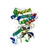
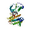
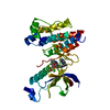
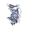
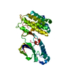
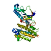
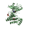
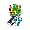
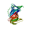
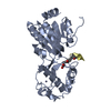
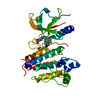
| Title | Crystal structure of c-MET bound by compound 3 | ||||||
 Components Components | Hepatocyte growth factor receptor | ||||||
 Keywords Keywords | TRANSFERASE / c-met / kinase / folded P-loop / inhibitor | ||||||
| Function / homology |  Function and homology information Function and homology informationhepatocyte growth factor receptor activity / Drug-mediated inhibition of MET activation / MET activates STAT3 / negative regulation of hydrogen peroxide-mediated programmed cell death / MET Receptor Activation / MET interacts with TNS proteins / endothelial cell morphogenesis / semaphorin receptor activity / MET receptor recycling / pancreas development ...hepatocyte growth factor receptor activity / Drug-mediated inhibition of MET activation / MET activates STAT3 / negative regulation of hydrogen peroxide-mediated programmed cell death / MET Receptor Activation / MET interacts with TNS proteins / endothelial cell morphogenesis / semaphorin receptor activity / MET receptor recycling / pancreas development / MET activates PTPN11 / hepatocyte growth factor receptor signaling pathway / MET activates RAP1 and RAC1 / Sema4D mediated inhibition of cell attachment and migration / positive regulation of endothelial cell chemotaxis / MET activates PI3K/AKT signaling / MET activates PTK2 signaling / branching morphogenesis of an epithelial tube / positive chemotaxis / semaphorin-plexin signaling pathway / Regulation of MITF-M-dependent genes involved in cell cycle and proliferation / MET activates RAS signaling / MECP2 regulates neuronal receptors and channels / cell surface receptor protein tyrosine kinase signaling pathway / molecular function activator activity / basal plasma membrane / negative regulation of autophagy / InlB-mediated entry of Listeria monocytogenes into host cell / liver development / excitatory postsynaptic potential / receptor protein-tyrosine kinase / Negative regulation of MET activity / Constitutive Signaling by Aberrant PI3K in Cancer / neuron differentiation / PIP3 activates AKT signaling / PI5P, PP2A and IER3 Regulate PI3K/AKT Signaling / RAF/MAP kinase cascade / protein tyrosine kinase activity / protein phosphatase binding / cell surface receptor signaling pathway / signaling receptor complex / postsynapse / cell surface / positive regulation of transcription by RNA polymerase II / extracellular region / ATP binding / membrane / identical protein binding / plasma membrane Similarity search - Function | ||||||
| Biological species |  Homo sapiens (human) Homo sapiens (human) | ||||||
| Method |  X-RAY DIFFRACTION / SYNCHROTRON / MOLECULAR REPLACEMENT / Resolution: 1.93 Å X-RAY DIFFRACTION / SYNCHROTRON / MOLECULAR REPLACEMENT / Resolution: 1.93 Å | ||||||
 Authors Authors | Collie, G.W. | ||||||
 Citation Citation | Journal: Acs Med.Chem.Lett. / Year: 2021 Title: Structural Basis for Targeting the Folded P-Loop Conformation of c-MET. Authors: Collie, G.W. / Michaelides, I.N. / Embrey, K. / Stubbs, C.J. / Borjesson, U. / Dale, I.L. / Snijder, A. / Barlind, L. / Song, K. / Khurana, P. / Phillips, C. / Storer, R.I. | ||||||
| History |
|
- Structure visualization
| Structure viewer | Molecule: MolmilJmol/JSmol |
|---|
- Downloads & links
-Download
| PDBx/mmCIF format | 7b3v.cif.gz | 75.9 KB | Display | PDBx/mmCIF format |
|---|---|---|---|---|
| PDB format | pdb7b3v.ent.gz | 53.9 KB | Display | PDB format |
| PDBx/mmJSON format | 7b3v.json.gz | Tree view | PDBx/mmJSON format | |
| Others |  Other downloads Other downloads |
-Validation report
| Arichive directory | https://data.pdbj.org/pub/pdb/validation_reports/b3/7b3vftp://data.pdbj.org/pub/pdb/validation_reports/b3/7b3v | HTTPS FTP |
|---|
-Related structure data
| Related structure data | 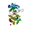 7b3qC 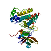 7b3tC 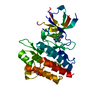 7b3wC  7b3zC 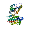 7b40C 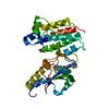 7b41C 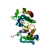 7b42C 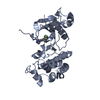 7b43C 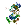 7b44C C: citing same article ( |
|---|---|
| Similar structure data |

-Links
 PDBj
PDBj


- Assembly
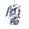
| Deposited unit | 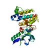
| ||||||||
|---|---|---|---|---|---|---|---|---|---|
| 1 |
| ||||||||
| Unit cell |
|
-Components
| #1: Protein | Mass: 33786.207 Da / Num. of mol.: 1 Source method: isolated from a genetically manipulated source Source: (gene. exp.) Homo sapiens (human) / Gene: MET / Production host:  References: UniProt: P08581, receptor protein-tyrosine kinase |
|---|---|
| #2: Chemical | ChemComp-SWB /   Mass: 252.271 Da / Num. of mol.: 1 / Source method: obtained synthetically / Formula: C14H12N4O / Feature type: SUBJECT OF INVESTIGATION Mass: 252.271 Da / Num. of mol.: 1 / Source method: obtained synthetically / Formula: C14H12N4O / Feature type: SUBJECT OF INVESTIGATION |
| #3: Water | ChemComp-HOH /  Mass: 18.015 Da / Num. of mol.: 222 / Source method: isolated from a natural source / Formula: H2O Mass: 18.015 Da / Num. of mol.: 222 / Source method: isolated from a natural source / Formula: H2O |
| Has ligand of interest | Y |
-Experimental details
-Experiment
| Experiment | Method: X-RAY DIFFRACTION / Number of used crystals: 1 |
|---|
- Sample preparation
| Crystal | Density Matthews: 2.6 Å3/Da / Density % sol: 52.73 % |
|---|---|
| Crystal grow | Temperature: 273 K / Method: vapor diffusion, sitting drop / Details: 25 % PEG3350, 0.2 M (NH4)2SO4, PCPT pH 5.5 |
-Data collection
| Diffraction | Mean temperature: 100 K / Serial crystal experiment: N |
|---|---|
| Diffraction source | Source: SYNCHROTRON / Site: Diamond  / Beamline: I03 / Wavelength: 0.97625 Å / Beamline: I03 / Wavelength: 0.97625 Å |
| Detector | Type: DECTRIS PILATUS 2M / Detector: PIXEL / Date: Jun 1, 2017 |
| Radiation | Protocol: SINGLE WAVELENGTH / Monochromatic (M) / Laue (L): M / Scattering type: x-ray |
| Radiation wavelength | Wavelength: 0.97625 Å / Relative weight: 1 |
| Reflection | Resolution: 1.93→28.71 Å / Num. obs: 26013 / % possible obs: 98 % / Redundancy: 3.2 % / CC1/2: 0.993 / Net I/σ(I): 18.8 |
| Reflection shell | Resolution: 1.93→1.96 Å / Num. unique obs: 1206 / CC1/2: 0.936 |
- Processing
| Software |
| ||||||||||||||||||||||||||||||||||||||||||||||||||||||||||||||||||||||||||||||||||||||||||||||||||||||||||||
|---|---|---|---|---|---|---|---|---|---|---|---|---|---|---|---|---|---|---|---|---|---|---|---|---|---|---|---|---|---|---|---|---|---|---|---|---|---|---|---|---|---|---|---|---|---|---|---|---|---|---|---|---|---|---|---|---|---|---|---|---|---|---|---|---|---|---|---|---|---|---|---|---|---|---|---|---|---|---|---|---|---|---|---|---|---|---|---|---|---|---|---|---|---|---|---|---|---|---|---|---|---|---|---|---|---|---|---|---|---|
| Refinement | Method to determine structure: MOLECULAR REPLACEMENT Starting model: internal Resolution: 1.93→22.61 Å / Cor.coef. Fo:Fc: 0.917 / Cor.coef. Fo:Fc free: 0.897 / Rfactor Rfree error: 0 / SU R Cruickshank DPI: 0.169 / Cross valid method: THROUGHOUT / σ(F): 0 / SU R Blow DPI: 0.177 / SU Rfree Blow DPI: 0.151 / SU Rfree Cruickshank DPI: 0.147
| ||||||||||||||||||||||||||||||||||||||||||||||||||||||||||||||||||||||||||||||||||||||||||||||||||||||||||||
| Displacement parameters | Biso max: 88.5 Å2 / Biso mean: 28.99 Å2 / Biso min: 11.28 Å2
| ||||||||||||||||||||||||||||||||||||||||||||||||||||||||||||||||||||||||||||||||||||||||||||||||||||||||||||
| Refine analyze | Luzzati coordinate error obs: 0.32 Å | ||||||||||||||||||||||||||||||||||||||||||||||||||||||||||||||||||||||||||||||||||||||||||||||||||||||||||||
| Refinement step | Cycle: final / Resolution: 1.93→22.61 Å
| ||||||||||||||||||||||||||||||||||||||||||||||||||||||||||||||||||||||||||||||||||||||||||||||||||||||||||||
| Refine LS restraints |
| ||||||||||||||||||||||||||||||||||||||||||||||||||||||||||||||||||||||||||||||||||||||||||||||||||||||||||||
| LS refinement shell | Resolution: 1.93→2.01 Å / Rfactor Rfree error: 0 / Total num. of bins used: 13
|
