 Movie
Movie Controller

[English] 日本語
 Yorodumi
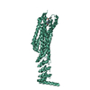
- PDB-3gpn: Structure of the non-trimeric form of the E113G PCNA mutant protein -
+ Open data

- Basic information
| Entry | Database: PDB / ID: 3gpn | ||||||
|---|---|---|---|---|---|---|---|
| Title | Structure of the non-trimeric form of the E113G PCNA mutant protein | ||||||
 Components Components | Proliferating cell nuclear antigen | ||||||
 Keywords Keywords | DNA BINDING PROTEIN / Replication / DNA damage / DNA repair / DNA replication / DNA-binding / Isopeptide bond / Nucleus / Ubl conjugation | ||||||
| Function / homology |  Function and homology information Function and homology informationMismatch repair (MMR) directed by MSH2:MSH6 (MutSalpha) / positive regulation of DNA metabolic process / Processive synthesis on the lagging strand / Removal of the Flap Intermediate / meiotic mismatch repair / Polymerase switching / maintenance of DNA trinucleotide repeats / SUMOylation of DNA replication proteins / establishment of mitotic sister chromatid cohesion / PCNA complex ...Mismatch repair (MMR) directed by MSH2:MSH6 (MutSalpha) / positive regulation of DNA metabolic process / Processive synthesis on the lagging strand / Removal of the Flap Intermediate / meiotic mismatch repair / Polymerase switching / maintenance of DNA trinucleotide repeats / SUMOylation of DNA replication proteins / establishment of mitotic sister chromatid cohesion / PCNA complex / lagging strand elongation / DNA damage tolerance / silent mating-type cassette heterochromatin formation / mitotic sister chromatid cohesion / error-free translesion synthesis / leading strand elongation / DNA polymerase processivity factor activity / subtelomeric heterochromatin formation / Dual incision in TC-NER / mismatch repair / translesion synthesis / positive regulation of DNA replication / positive regulation of DNA repair / replication fork / nucleotide-excision repair / mitotic cell cycle / chromosome, telomeric region / DNA binding / identical protein binding / nucleus Similarity search - Function | ||||||
| Biological species |  | ||||||
| Method |  X-RAY DIFFRACTION / SYNCHROTRON / MOLECULAR REPLACEMENT / Resolution: 2.5 Å X-RAY DIFFRACTION / SYNCHROTRON / MOLECULAR REPLACEMENT / Resolution: 2.5 Å | ||||||
 Authors Authors | Freudenthal, B.D. / Gakhar, L. / Ramaswamy, S. / Washington, M.T. | ||||||
 Citation Citation | Journal: Acta Crystallogr.,Sect.D / Year: 2009 Title: A charged residue at the subunit interface of PCNA promotes trimer formation by destabilizing alternate subunit interactions. Authors: Freudenthal, B.D. / Gakhar, L. / Ramaswamy, S. / Washington, M.T. | ||||||
| History |
|
- Structure visualization
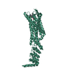
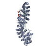
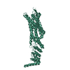
| Structure viewer | Molecule: MolmilJmol/JSmol |
|---|
- Downloads & links
-Download
| PDBx/mmCIF format | 3gpn.cif.gz | 60 KB | Display | PDBx/mmCIF format |
|---|---|---|---|---|
| PDB format | pdb3gpn.ent.gz | 44.3 KB | Display | PDB format |
| PDBx/mmJSON format | 3gpn.json.gz | Tree view | PDBx/mmJSON format | |
| Others |  Other downloads Other downloads |
-Validation report
| Arichive directory | https://data.pdbj.org/pub/pdb/validation_reports/gp/3gpnftp://data.pdbj.org/pub/pdb/validation_reports/gp/3gpn | HTTPS FTP |
|---|
-Related structure data
| Related structure data |  3gpmC 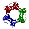 1plqS C: citing same article ( S: Starting model for refinement |
|---|---|
| Similar structure data |


-Links
 PDBj
PDBj


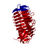
- Assembly
| Deposited unit | 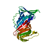
| ||||||||
|---|---|---|---|---|---|---|---|---|---|
| 1 | 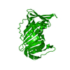
| ||||||||
| Unit cell |
|
-Components
| #1: Protein | Mass: 28871.988 Da / Num. of mol.: 1 / Mutation: E113G Source method: isolated from a genetically manipulated source Source: (gene. exp.) Gene: POL30, YBR0811, YBR088C / Plasmid: Pet11-a / Production host:  |
|---|
-Experimental details
-Experiment
| Experiment | Method: X-RAY DIFFRACTION / Number of used crystals: 1 |
|---|
- Sample preparation
| Crystal | Density Matthews: 3.97 Å3/Da / Density % sol: 68.99 % |
|---|---|
| Crystal grow | Temperature: 291 K / Method: vapor diffusion, hanging drop / pH: 5.8 Details: 1.6 M ammonium sulfate and 0.1 M sodium Citrate, pH 5.8, VAPOR DIFFUSION, HANGING DROP, temperature 291K |
-Data collection
| Diffraction | Mean temperature: 100 K |
|---|---|
| Diffraction source | Source: SYNCHROTRON / Site: ALS  / Beamline: 4.2.2 / Wavelength: 1.072 Å / Beamline: 4.2.2 / Wavelength: 1.072 Å |
| Detector | Type: NOIR-1 / Detector: CCD / Date: Feb 4, 2008 / Details: saggital focusing mirrors |
| Radiation | Monochromator: Sagitally focused Si(111) / Protocol: SINGLE WAVELENGTH / Monochromatic (M) / Laue (L): M / Scattering type: x-ray |
| Radiation wavelength | Wavelength: 1.072 Å / Relative weight: 1 |
| Reflection | Resolution: 2.5→81.38 Å / Num. all: 15940 / Num. obs: 14864 / % possible obs: 98.18 % / Redundancy: 4.75 % / Biso Wilson estimate: 42.619 Å2 / Rsym value: 0.09 / Net I/σ(I): 11.4 |
| Reflection shell | Resolution: 2.5→2.565 Å / Redundancy: 4.75 % / Rmerge(I) obs: 0.365 / Mean I/σ(I) obs: 3.2 / Num. unique all: 1041 / Rsym value: 0.365 / % possible all: 96.79 |
- Processing
| Software |
| ||||||||||||||||||||||||||||||||||||||||||||||||||||||||||||||||||||||||||||||||||||||||||
|---|---|---|---|---|---|---|---|---|---|---|---|---|---|---|---|---|---|---|---|---|---|---|---|---|---|---|---|---|---|---|---|---|---|---|---|---|---|---|---|---|---|---|---|---|---|---|---|---|---|---|---|---|---|---|---|---|---|---|---|---|---|---|---|---|---|---|---|---|---|---|---|---|---|---|---|---|---|---|---|---|---|---|---|---|---|---|---|---|---|---|---|
| Refinement | Method to determine structure: MOLECULAR REPLACEMENT Starting model: 1PLQ Resolution: 2.5→81.38 Å / Cor.coef. Fo:Fc: 0.924 / Cor.coef. Fo:Fc free: 0.904 / SU B: 10.022 / SU ML: 0.221 / Isotropic thermal model: isotropic / Cross valid method: THROUGHOUT / σ(F): 0.02 / ESU R: 0.32 / ESU R Free: 0.255 / Stereochemistry target values: MAXIMUM LIKELIHOOD / Details: HYDROGENS HAVE BEEN ADDED IN THE RIDING POSITIONS
| ||||||||||||||||||||||||||||||||||||||||||||||||||||||||||||||||||||||||||||||||||||||||||
| Solvent computation | Ion probe radii: 0.8 Å / Shrinkage radii: 0.8 Å / VDW probe radii: 1.2 Å / Solvent model: MASK | ||||||||||||||||||||||||||||||||||||||||||||||||||||||||||||||||||||||||||||||||||||||||||
| Displacement parameters | Biso mean: 42.619 Å2
| ||||||||||||||||||||||||||||||||||||||||||||||||||||||||||||||||||||||||||||||||||||||||||
| Refinement step | Cycle: LAST / Resolution: 2.5→81.38 Å
| ||||||||||||||||||||||||||||||||||||||||||||||||||||||||||||||||||||||||||||||||||||||||||
| Refine LS restraints |
| ||||||||||||||||||||||||||||||||||||||||||||||||||||||||||||||||||||||||||||||||||||||||||
| LS refinement shell | Refine-ID: X-RAY DIFFRACTION / Total num. of bins used: 20
|
