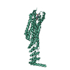
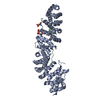
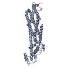
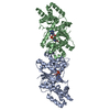
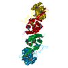
Entry Database : PDB / ID : 3ouxTitle Structure of beta-catenin with phosphorylated Lef-1 Catenin beta-1 Lymphoid enhancer-binding factor 1 Keywords / / / / / Function / homology Function Domain/homology Component
/ / / / / / / / / / / / / / / / / / / / / / / / / / / / / / / / / / / / / / / / / / / / / / / / / / / / / / / / / / / / / / / / / / / / / / / / / / / / / / / / / / / / / / / / / / / / / / / / / / / / / / / / / / / / / / / / / / / / / / / / / / / / / / / / / / / / / / / / / / / / Biological species Mus musculus (house mouse)Method / / / Resolution : 2.4 Å Authors Weis, W.I. / Sun, J. Journal : J.Mol.Biol. / Year : 2011Title : Biochemical and structural characterization of beta-catenin interactions with nonphosphorylated and CK2-phosphorylated Lef-1.Authors : Sun, J. / Weis, W.I. History Deposition Sep 15, 2010 Deposition site / Processing site Revision 1.0 Nov 24, 2010 Provider / Type Revision 1.1 Jul 13, 2011 Group Revision 1.2 Dec 2, 2015 Group Revision 1.3 Dec 9, 2015 Group Revision 1.4 Sep 6, 2023 Group / Database references / Refinement descriptionCategory chem_comp_atom / chem_comp_bond ... chem_comp_atom / chem_comp_bond / database_2 / pdbx_initial_refinement_model / struct_ref_seq_dif Item / _database_2.pdbx_database_accession / _struct_ref_seq_dif.detailsRevision 1.5 Oct 30, 2024 Group / Category / pdbx_modification_feature
Show all Show less
 Movie
Movie Controller


 Open data
 Basic information
 Components
 Keywords
 Function and homology information
 X-RAY DIFFRACTION /
  Authors
 Citation
 Structure visualization
 Downloads & links
 Other downloads
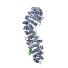


 PDBj
PDBj


 Assembly
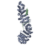

 Mass: 18.015 Da / Num. of mol.: 97 / Source method: isolated from a natural source / Formula: H2O
 Sample preparation
 / Beamline: BL9-1 / Wavelength: 0.97945 Å
 Processing