 Movie
Movie Controller

[English] 日本語
 Yorodumi
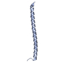
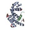
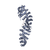
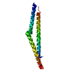
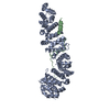
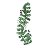
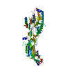
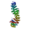
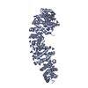
- PDB-1v18: The crystal structure of beta-catenin armadillo repeat complexed ... -
+ Open data

- Basic information
| Entry | Database: PDB / ID: 1v18 | ||||||
|---|---|---|---|---|---|---|---|
| Title | The crystal structure of beta-catenin armadillo repeat complexed with a phosphorylated APC 20mer repeat. | ||||||
 Components Components |
| ||||||
 Keywords Keywords | SIGNALING PROTEIN / SIGNALLING COMPLEX / WNT SIGNAL / BETA-CATENIN DEGRADATION COMPLEX / CELL ADHESION / TRANSCRIPTION / TRANSCRIPTION REGULATION | ||||||
| Function / homology |  Function and homology information Function and homology informationmetanephros morphogenesis / lung cell differentiation / lung induction / epicardium-derived cardiac vascular smooth muscle cell differentiation / positive regulation of branching involved in lung morphogenesis / renal vesicle formation / renal inner medulla development / renal outer medulla development / nephron tubule formation / mesenchyme morphogenesis ...metanephros morphogenesis / lung cell differentiation / lung induction / epicardium-derived cardiac vascular smooth muscle cell differentiation / positive regulation of branching involved in lung morphogenesis / renal vesicle formation / renal inner medulla development / renal outer medulla development / nephron tubule formation / mesenchyme morphogenesis / genitalia morphogenesis / RUNX3 regulates WNT signaling / neural plate development / glial cell fate determination / Regulation of CDH11 function / oviduct development / cardiac vascular smooth muscle cell differentiation / regulation of secondary heart field cardioblast proliferation / Regulation of MITF-M-dependent genes involved in cell cycle and proliferation / central nervous system vasculogenesis / trachea morphogenesis / negative regulation of mesenchymal to epithelial transition involved in metanephros morphogenesis / smooth muscle cell differentiation / Beta-catenin phosphorylation cascade / APC truncation mutants are not K63 polyubiquitinated / positive regulation of epithelial cell proliferation involved in prostate gland development / Apoptotic cleavage of cell adhesion proteins / endoderm formation / proximal/distal pattern formation / hair cycle process / embryonic foregut morphogenesis / ectoderm development / Disassembly of the destruction complex and recruitment of AXIN to the membrane / embryonic axis specification / LRR FLII-interacting protein 1 (LRRFIP1) activates type I IFN production / lens morphogenesis in camera-type eye / regulation of epithelial cell differentiation / positive regulation of epithelial cell differentiation / Formation of the beta-catenin:TCF transactivating complex / dorsal/ventral axis specification / Regulation of CDH1 posttranslational processing and trafficking to plasma membrane / epithelial cell differentiation involved in prostate gland development / positive regulation of heparan sulfate proteoglycan biosynthetic process / cranial ganglion development / embryonic skeletal limb joint morphogenesis / astrocyte-dopaminergic neuron signaling / VEGFR2 mediated vascular permeability / beta-catenin-TCF7L2 complex / regulation of nephron tubule epithelial cell differentiation / gastrulation with mouth forming second / negative regulation of cyclin-dependent protein serine/threonine kinase activity / regulation of timing of anagen / endodermal cell fate commitment / Schwann cell proliferation / negative regulation of mitotic cell cycle, embryonic / establishment of blood-retinal barrier / TCF dependent signaling in response to WNT / regulation of centriole-centriole cohesion / Adherens junctions interactions / positive regulation of fibroblast growth factor receptor signaling pathway / Deactivation of the beta-catenin transactivating complex / layer formation in cerebral cortex / regulation of centromeric sister chromatid cohesion / morphogenesis of embryonic epithelium / mesenchymal cell proliferation involved in lung development / animal organ development / lung-associated mesenchyme development / negative regulation of cell cycle G1/S phase transition / positive regulation of endothelial cell differentiation / mesenchyme development / Ca2+ pathway / fungiform papilla formation / positive regulation of determination of dorsal identity / Scrib-APC-beta-catenin complex / establishment of blood-brain barrier / beta-catenin-TCF complex / RHO GTPases activate IQGAPs / oocyte development / positive regulation of skeletal muscle tissue development / embryonic hindlimb morphogenesis / synaptic vesicle clustering / Degradation of beta-catenin by the destruction complex / Regulation of CDH1 Function / male genitalia development / Degradation of CDH1 / embryonic forelimb morphogenesis / dorsal root ganglion development / ventricular compact myocardium morphogenesis / gamma-catenin binding / regulation of attachment of spindle microtubules to kinetochore / endothelial tube morphogenesis / hindbrain development / delta-catenin binding / mesenchymal to epithelial transition / sympathetic ganglion development / negative regulation of oligodendrocyte differentiation / tissue homeostasis / cranial skeletal system development / presynaptic active zone cytoplasmic component / regulation of protein localization to cell surface Similarity search - Function | ||||||
| Biological species |   HOMO SAPIENS (human) HOMO SAPIENS (human) | ||||||
| Method |  X-RAY DIFFRACTION / SYNCHROTRON / MOLECULAR REPLACEMENT / Resolution: 2.1 Å X-RAY DIFFRACTION / SYNCHROTRON / MOLECULAR REPLACEMENT / Resolution: 2.1 Å | ||||||
 Authors Authors | Ha, N.-C. / Weis, W.I. | ||||||
 Citation Citation | Journal: Mol.Cell / Year: 2004 Title: Mechanism of Phosphorylation-Dependent Binding of Apc to Beta-Catenin and its Role in Beta-Catenin Degradation Authors: Ha, N.-C. / Tonozuka, T. / Stamos, J.L. / Weis, W.I. | ||||||
| History |
|
- Structure visualization
| Structure viewer | Molecule: MolmilJmol/JSmol |
|---|
- Downloads & links
-Download
| PDBx/mmCIF format | 1v18.cif.gz | 121.5 KB | Display | PDBx/mmCIF format |
|---|---|---|---|---|
| PDB format | pdb1v18.ent.gz | 92.8 KB | Display | PDB format |
| PDBx/mmJSON format | 1v18.json.gz | Tree view | PDBx/mmJSON format | |
| Others |  Other downloads Other downloads |
-Validation report
| Arichive directory | https://data.pdbj.org/pub/pdb/validation_reports/v1/1v18ftp://data.pdbj.org/pub/pdb/validation_reports/v1/1v18 | HTTPS FTP |
|---|
-Related structure data
| Related structure data | 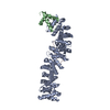 1t08C  1i7wS S: Starting model for refinement C: citing same article ( |
|---|---|
| Similar structure data |


-Links
 PDBj
PDBj


- Assembly
| Deposited unit | 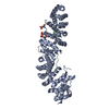
| ||||||||
|---|---|---|---|---|---|---|---|---|---|
| 1 |
| ||||||||
| Unit cell |
|
-Components
| #1: Protein | Mass: 58844.117 Da / Num. of mol.: 1 Fragment: ARMADILLO REPEAT, REPEAT 3 OF APC, RESIDUES 134-671 Source method: isolated from a genetically manipulated source Details: 134-671 OF BETA-CATENIN AND 1484-1528 OF HUMAN APC(ADENOMATOUS POLYPOSIS COLI) PHOSPHORYLATED BY CK1 AND GSK-3BETA Source: (gene. exp.)  |
|---|---|
| #2: Protein/peptide | Mass: 5414.617 Da / Num. of mol.: 1 / Fragment: RESIDUES 1482-1528 Source method: isolated from a genetically manipulated source Details: 134-671 OF BETA-CATENIN AND 1484-1528 OF HUMAN APC(ADENOMATOUS POLYPOSIS COLI) PHOSPHORYLATED BY CK1 AND GSK-3BETA Source: (gene. exp.) HOMO SAPIENS (human) / Plasmid: PPROEXHTA / Production host: |
| #3: Water | ChemComp-HOH /  Mass: 18.015 Da / Num. of mol.: 235 / Source method: isolated from a natural source / Formula: H2O Mass: 18.015 Da / Num. of mol.: 235 / Source method: isolated from a natural source / Formula: H2O |
| Has protein modification | Y |
-Experimental details
-Experiment
| Experiment | Method: X-RAY DIFFRACTION / Number of used crystals: 1 |
|---|
- Sample preparation
| Crystal | Density Matthews: 2.19 Å3/Da / Density % sol: 43.76 % |
|---|---|
| Crystal grow | pH: 6.5 / Details: pH 6.50 |
-Data collection
| Diffraction | Mean temperature: 100 K |
|---|---|
| Diffraction source | Source: SYNCHROTRON / Site: ALS  / Beamline: 8.2.2 / Wavelength: 1 / Beamline: 8.2.2 / Wavelength: 1 |
| Detector | Type: ADSC CCD / Detector: CCD |
| Radiation | Protocol: SINGLE WAVELENGTH / Monochromatic (M) / Laue (L): M / Scattering type: x-ray |
| Radiation wavelength | Wavelength: 1 Å / Relative weight: 1 |
| Reflection | Resolution: 2.1→50 Å / Num. obs: 31262 / % possible obs: 97.4 % / Redundancy: 7 % / Biso Wilson estimate: 23.7 Å2 / Rmerge(I) obs: 0.069 / Net I/σ(I): 37 |
| Reflection shell | Resolution: 2.1→2.18 Å / Redundancy: 2.6 % / Rmerge(I) obs: 0.313 / Mean I/σ(I) obs: 3.6 / % possible all: 85.5 |
- Processing
| Software |
| ||||||||||||||||||||||||||||||||||||||||||||||||||||||||||||||||||||||||||||||||
|---|---|---|---|---|---|---|---|---|---|---|---|---|---|---|---|---|---|---|---|---|---|---|---|---|---|---|---|---|---|---|---|---|---|---|---|---|---|---|---|---|---|---|---|---|---|---|---|---|---|---|---|---|---|---|---|---|---|---|---|---|---|---|---|---|---|---|---|---|---|---|---|---|---|---|---|---|---|---|---|---|---|
| Refinement | Method to determine structure: MOLECULAR REPLACEMENT Starting model: PDB ENTRY 1I7W Resolution: 2.1→50 Å / Rfactor Rfree error: 0.005 / Data cutoff high absF: 10000 / Isotropic thermal model: RESTRAINED / Cross valid method: THROUGHOUT / σ(F): 0
| ||||||||||||||||||||||||||||||||||||||||||||||||||||||||||||||||||||||||||||||||
| Solvent computation | Solvent model: FLAT MODEL / Bsol: 61.5715 Å2 / ksol: 0.376423 e/Å3 | ||||||||||||||||||||||||||||||||||||||||||||||||||||||||||||||||||||||||||||||||
| Displacement parameters | Biso mean: 47.6 Å2
| ||||||||||||||||||||||||||||||||||||||||||||||||||||||||||||||||||||||||||||||||
| Refine analyze |
| ||||||||||||||||||||||||||||||||||||||||||||||||||||||||||||||||||||||||||||||||
| Refinement step | Cycle: LAST / Resolution: 2.1→50 Å
| ||||||||||||||||||||||||||||||||||||||||||||||||||||||||||||||||||||||||||||||||
| Refine LS restraints |
| ||||||||||||||||||||||||||||||||||||||||||||||||||||||||||||||||||||||||||||||||
| LS refinement shell | Resolution: 2.1→2.23 Å / Rfactor Rfree error: 0.015 / Total num. of bins used: 6
| ||||||||||||||||||||||||||||||||||||||||||||||||||||||||||||||||||||||||||||||||
| Xplor file |
|
