 Movie
Movie Controller

[English] 日本語
 Yorodumi
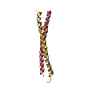
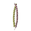
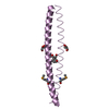
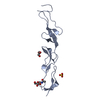
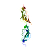
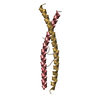
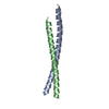
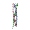
- PDB-1m5i: Crystal Structure of the coiled coil region 129-250 of the tumor ... -
+ Open data

- Basic information
| Entry | Database: PDB / ID: 1m5i | ||||||
|---|---|---|---|---|---|---|---|
| Title | Crystal Structure of the coiled coil region 129-250 of the tumor suppressor gene product APC | ||||||
 Components Components | APC protein | ||||||
 Keywords Keywords | ANTITUMOR PROTEIN / coiled-coil | ||||||
| Function / homology |  Function and homology information Function and homology informationAPC truncation mutants are not K63 polyubiquitinated / negative regulation of cell cycle G1/S phase transition / negative regulation of cyclin-dependent protein serine/threonine kinase activity / gamma-catenin binding / regulation of attachment of spindle microtubules to kinetochore / pattern specification process / positive regulation of pseudopodium assembly / positive regulation of protein localization to centrosome / negative regulation of microtubule depolymerization / bicellular tight junction assembly ...APC truncation mutants are not K63 polyubiquitinated / negative regulation of cell cycle G1/S phase transition / negative regulation of cyclin-dependent protein serine/threonine kinase activity / gamma-catenin binding / regulation of attachment of spindle microtubules to kinetochore / pattern specification process / positive regulation of pseudopodium assembly / positive regulation of protein localization to centrosome / negative regulation of microtubule depolymerization / bicellular tight junction assembly / heart valve development / beta-catenin destruction complex / catenin complex / APC truncation mutants have impaired AXIN binding / AXIN missense mutants destabilize the destruction complex / Truncations of AMER1 destabilize the destruction complex / microtubule plus-end binding / protein kinase regulator activity / Beta-catenin phosphorylation cascade / Signaling by GSK3beta mutants / CTNNB1 S33 mutants aren't phosphorylated / CTNNB1 S37 mutants aren't phosphorylated / CTNNB1 S45 mutants aren't phosphorylated / CTNNB1 T41 mutants aren't phosphorylated / cell fate specification / Wnt signalosome / regulation of microtubule-based process / Disassembly of the destruction complex and recruitment of AXIN to the membrane / endocardial cushion morphogenesis / Apoptotic cleavage of cellular proteins / mitotic spindle assembly checkpoint signaling / negative regulation of G1/S transition of mitotic cell cycle / regulation of microtubule-based movement / mitotic cytokinesis / dynein complex binding / lateral plasma membrane / bicellular tight junction / adherens junction / Deactivation of the beta-catenin transactivating complex / negative regulation of canonical Wnt signaling pathway / beta-catenin binding / kinetochore / Degradation of beta-catenin by the destruction complex / Wnt signaling pathway / ruffle membrane / positive regulation of protein catabolic process / insulin receptor signaling pathway / Ovarian tumor domain proteases / nervous system development / cell migration / lamellipodium / positive regulation of cold-induced thermogenesis / protein-containing complex assembly / microtubule binding / microtubule / proteasome-mediated ubiquitin-dependent protein catabolic process / cell adhesion / positive regulation of cell migration / positive regulation of apoptotic process / negative regulation of cell population proliferation / centrosome / ubiquitin protein ligase binding / DNA damage response / protein kinase binding / perinuclear region of cytoplasm / Golgi apparatus / nucleoplasm / nucleus / plasma membrane / cytosol / cytoplasm Similarity search - Function | ||||||
| Biological species |  Homo sapiens (human) Homo sapiens (human) | ||||||
| Method |  X-RAY DIFFRACTION / SYNCHROTRON / SAD / Resolution: 2 Å X-RAY DIFFRACTION / SYNCHROTRON / SAD / Resolution: 2 Å | ||||||
 Authors Authors | Tickenbrock, L. / Cramer, J. / Vetter, I.R. / Mueller, O. | ||||||
 Citation Citation | Journal: J.Biol.Chem. / Year: 2002 Title: The coiled coil region (amino acids 129-250) of the tumor suppressor protein adenomatous polyposis coli (APC). Its structure and its interaction with chromosome maintenance region 1 (Crm-1). Authors: Tickenbrock, L. / Cramer, J. / Vetter, I.R. / Muller, O. #1: Journal: HUM.MOL.GENET. / Year: 2001Title: The ABC of APC Authors: Fearnhead, N.S. / Britton, M.P. / Bodmer, W.F. | ||||||
| History |
|
- Structure visualization
| Structure viewer | Molecule: MolmilJmol/JSmol |
|---|
- Downloads & links
-Download
| PDBx/mmCIF format | 1m5i.cif.gz | 34.6 KB | Display | PDBx/mmCIF format |
|---|---|---|---|---|
| PDB format | pdb1m5i.ent.gz | 23.8 KB | Display | PDB format |
| PDBx/mmJSON format | 1m5i.json.gz | Tree view | PDBx/mmJSON format | |
| Others |  Other downloads Other downloads |
-Validation report
| Arichive directory | https://data.pdbj.org/pub/pdb/validation_reports/m5/1m5iftp://data.pdbj.org/pub/pdb/validation_reports/m5/1m5i | HTTPS FTP |
|---|
-Related structure data
| Similar structure data |
|---|


-Links
 PDBj
PDBj


- Assembly
| Deposited unit | 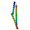
| |||||||||
|---|---|---|---|---|---|---|---|---|---|---|
| 1 |
| |||||||||
| Unit cell |
| |||||||||
| Components on special symmetry positions |
|
-Components
| #1: Protein | Mass: 14952.779 Da / Num. of mol.: 1 / Fragment: coiled-coil region Source method: isolated from a genetically manipulated source Source: (gene. exp.) Homo sapiens (human) / Gene: APC / Plasmid: pGEX-6P-2 / Species (production host): Escherichia coli / Production host:  |
|---|---|
| #2: Water | ChemComp-HOH /  Mass: 18.015 Da / Num. of mol.: 56 / Source method: isolated from a natural source / Formula: H2O Mass: 18.015 Da / Num. of mol.: 56 / Source method: isolated from a natural source / Formula: H2O |
-Experimental details
-Experiment
| Experiment | Method: X-RAY DIFFRACTION / Number of used crystals: 1 |
|---|
- Sample preparation
| Crystal | Density Matthews: 2.32 Å3/Da / Density % sol: 46.95 % | ||||||||||||||||||||||||||||||
|---|---|---|---|---|---|---|---|---|---|---|---|---|---|---|---|---|---|---|---|---|---|---|---|---|---|---|---|---|---|---|---|
| Crystal grow | Temperature: 277 K / Method: vapor diffusion, hanging drop Details: 16% PEG 3350, 100mM potassium iodide, 10mM strontium chloride, VAPOR DIFFUSION, HANGING DROP, temperature 277K | ||||||||||||||||||||||||||||||
| Crystal grow | *PLUS Temperature: 4 ℃ | ||||||||||||||||||||||||||||||
| Components of the solutions | *PLUS
|
-Data collection
| Diffraction | Mean temperature: 100 K |
|---|---|
| Diffraction source | Source: SYNCHROTRON / Site: ESRF  / Beamline: ID14-1 / Wavelength: 0.934 Å / Beamline: ID14-1 / Wavelength: 0.934 Å |
| Detector | Type: MARRESEARCH / Detector: CCD / Date: Feb 4, 2001 / Details: mirrors |
| Radiation | Monochromator: SAGITALLY FOCUSED GE(220) / Protocol: SINGLE WAVELENGTH / Monochromatic (M) / Laue (L): M / Scattering type: x-ray |
| Radiation wavelength | Wavelength: 0.934 Å / Relative weight: 1 |
| Reflection | Resolution: 2→19.94 Å / Num. all: 10548 / Num. obs: 10548 / % possible obs: 98.7 % / Observed criterion σ(F): 0 / Observed criterion σ(I): 0 / Redundancy: 6.3 % / Biso Wilson estimate: 38.6 Å2 / Rmerge(I) obs: 0.05 / Rsym value: 0.067 / Net I/σ(I): 18.8 |
| Reflection shell | Resolution: 2→2.1 Å / Redundancy: 5.3 % / Rmerge(I) obs: 0.127 / Mean I/σ(I) obs: 7 / Num. unique all: 2317 / Rsym value: 0.216 / % possible all: 98.8 |
| Reflection | *PLUS Lowest resolution: 40 Å / Num. obs: 9685 / % possible obs: 98.6 % / Num. measured all: 65629 / Rmerge(I) obs: 0.067 |
| Reflection shell | *PLUS % possible obs: 98.8 % / Mean I/σ(I) obs: 6.3 |
- Processing
| Software |
| |||||||||||||||||||||||||
|---|---|---|---|---|---|---|---|---|---|---|---|---|---|---|---|---|---|---|---|---|---|---|---|---|---|---|
| Refinement | Method to determine structure: SAD / Resolution: 2→20 Å / Isotropic thermal model: overall / Cross valid method: THROUGHOUT / σ(F): 0 / Stereochemistry target values: Engh & Huber
| |||||||||||||||||||||||||
| Displacement parameters | Biso mean: 42.8 Å2 | |||||||||||||||||||||||||
| Refine analyze |
| |||||||||||||||||||||||||
| Refinement step | Cycle: LAST / Resolution: 2→20 Å
| |||||||||||||||||||||||||
| Refine LS restraints |
| |||||||||||||||||||||||||
| LS refinement shell | Resolution: 2→2.1 Å / Rfactor Rfree error: 0.027
| |||||||||||||||||||||||||
| Refinement | *PLUS Num. reflection obs: 10548 / % reflection Rfree: 10 % | |||||||||||||||||||||||||
| Solvent computation | *PLUS | |||||||||||||||||||||||||
| Displacement parameters | *PLUS |
