 Movie
Movie Controller

[English] 日本語
 Yorodumi
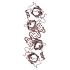
- PDB-4ywt: Crystal structure of full-length glypican-1 core protein after co... -
+ Open data

- Basic information
| Entry | Database: PDB / ID: 4ywt | ||||||
|---|---|---|---|---|---|---|---|
| Title | Crystal structure of full-length glypican-1 core protein after controlled crystal dehydration to 87% relative humidity | ||||||
 Components Components | Glypican-1 | ||||||
 Keywords Keywords | MEMBRANE PROTEIN / glypican-1 / diffraction quality / controlled dehydration / HC1b | ||||||
| Function / homology |  Function and homology information Function and homology informationpositive regulation of skeletal muscle cell differentiation / regulation of protein localization to membrane / Defective B3GALT6 causes EDSP2 and SEMDJL1 / myelin assembly / Defective B4GALT7 causes EDS, progeroid type / Defective B3GAT3 causes JDSSDHD / Defective EXT2 causes exostoses 2 / Defective EXT1 causes exostoses 1, TRPS2 and CHDS / Schwann cell differentiation / Glycosaminoglycan-protein linkage region biosynthesis ...positive regulation of skeletal muscle cell differentiation / regulation of protein localization to membrane / Defective B3GALT6 causes EDSP2 and SEMDJL1 / myelin assembly / Defective B4GALT7 causes EDS, progeroid type / Defective B3GAT3 causes JDSSDHD / Defective EXT2 causes exostoses 2 / Defective EXT1 causes exostoses 1, TRPS2 and CHDS / Schwann cell differentiation / Glycosaminoglycan-protein linkage region biosynthesis / HS-GAG biosynthesis / negative regulation of fibroblast growth factor receptor signaling pathway / heparan sulfate proteoglycan catabolic process / HS-GAG degradation / Signaling by ROBO receptors / RSV-host interactions / Respiratory syncytial virus (RSV) attachment and entry / fibroblast growth factor binding / Retinoid metabolism and transport / side of membrane / laminin binding / lysosomal lumen / Cell surface interactions at the vascular wall / Golgi lumen / cell migration / extracellular matrix / Attachment and Entry / endosome / membrane raft / copper ion binding / synapse / cell surface / : / extracellular exosome / extracellular region / nucleoplasm / plasma membrane / cytosol Similarity search - Function | ||||||
| Biological species |  Homo sapiens (human) Homo sapiens (human) | ||||||
| Method |  X-RAY DIFFRACTION / SYNCHROTRON / MOLECULAR REPLACEMENT / Resolution: 2.38 Å X-RAY DIFFRACTION / SYNCHROTRON / MOLECULAR REPLACEMENT / Resolution: 2.38 Å | ||||||
 Authors Authors | Awad, W. / Mani, K. / Logan, D.T. | ||||||
 Citation Citation | Journal: J.Biol.Chem. / Year: 2015 Title: Structural Aspects of N-Glycosylations and the C-terminal Region in Human Glypican-1. Authors: Awad, W. / Adamczyk, B. / Ornros, J. / Karlsson, N.G. / Mani, K. / Logan, D.T. #1: Journal: Acta Crystallogr. D Biol. Crystallogr. / Year: 2013Title: Improvements in the order, isotropy and electron density of glypican-1 crystals by controlled dehydration. Authors: Awad, W. / Svensson Birkedal, G. / Thunnissen, M.M. / Mani, K. / Logan, D.T. #2: Journal: J. Biol. Chem. / Year: 2012Title: Crystal structure of N-glycosylated human glypican-1 core protein: structure of two loops evolutionarily conserved in vertebrate glypican-1. Authors: Svensson, G. / Awad, W. / Hakansson, M. / Mani, K. / Logan, D.T. | ||||||
| History |
|
- Structure visualization
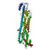
| Structure viewer | Molecule: MolmilJmol/JSmol |
|---|
- Downloads & links
-Download
| PDBx/mmCIF format | 4ywt.cif.gz | 659.4 KB | Display | PDBx/mmCIF format |
|---|---|---|---|---|
| PDB format | pdb4ywt.ent.gz | 540.8 KB | Display | PDB format |
| PDBx/mmJSON format | 4ywt.json.gz | Tree view | PDBx/mmJSON format | |
| Others |  Other downloads Other downloads |
-Validation report
| Arichive directory | https://data.pdbj.org/pub/pdb/validation_reports/yw/4ywtftp://data.pdbj.org/pub/pdb/validation_reports/yw/4ywt | HTTPS FTP |
|---|
-Related structure data
| Related structure data |  4bweS S: Starting model for refinement |
|---|---|
| Similar structure data |


-Links
 PDBj
PDBj


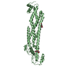
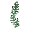
- Assembly
| Deposited unit | 
| ||||||||
|---|---|---|---|---|---|---|---|---|---|
| 1 | 
| ||||||||
| 2 | 
| ||||||||
| 3 | 
| ||||||||
| 4 | 
| ||||||||
| Unit cell |
|
-Components
| #1: Protein | Mass: 58506.973 Da / Num. of mol.: 4 / Fragment: UNP residues 24-527 / Mutation: S486A, S488A and S490A Source method: isolated from a genetically manipulated source Source: (gene. exp.) Homo sapiens (human) / Gene: GPC1 / Cell line (production host): HEK293 / Production host: Homo sapiens (human) / References: UniProt: P35052#2: Sugar | ChemComp-NAG /   Type: D-saccharide, beta linking / Mass: 221.208 Da / Num. of mol.: 4 Type: D-saccharide, beta linking / Mass: 221.208 Da / Num. of mol.: 4Source method: isolated from a genetically manipulated source Formula: C8H15NO6 #3: Chemical | ChemComp-CA /   Mass: 40.078 Da / Num. of mol.: 9 / Source method: obtained synthetically / Formula: Ca Mass: 40.078 Da / Num. of mol.: 9 / Source method: obtained synthetically / Formula: Ca#4: Water | ChemComp-HOH / |  Mass: 18.015 Da / Num. of mol.: 284 / Source method: isolated from a natural source / Formula: H2O Mass: 18.015 Da / Num. of mol.: 284 / Source method: isolated from a natural source / Formula: H2OHas protein modification | Y | |
|---|
-Experimental details
-Experiment
| Experiment | Method: X-RAY DIFFRACTION / Number of used crystals: 1 |
|---|
- Sample preparation
| Crystal | Density Matthews: 2.29 Å3/Da / Density % sol: 46.31 % |
|---|---|
| Crystal grow | Temperature: 298 K / Method: vapor diffusion, sitting drop / pH: 8 / Details: PEG 6000, Tris-HCl and CaCl2 / PH range: 8 |
-Data collection
| Diffraction | Mean temperature: 100 K |
|---|---|
| Diffraction source | Source: SYNCHROTRON / Site: MAX II  / Beamline: I911-3 / Wavelength: 1 Å / Beamline: I911-3 / Wavelength: 1 Å |
| Detector | Type: MARMOSAIC 225 mm CCD / Detector: CCD / Date: May 8, 2014 |
| Radiation | Monochromator: double crystal, Si[111] / Protocol: SINGLE WAVELENGTH / Monochromatic (M) / Laue (L): M / Scattering type: x-ray |
| Radiation wavelength | Wavelength: 1 Å / Relative weight: 1 |
| Reflection | Resolution: 2.38→43.22 Å / Num. obs: 82475 / % possible obs: 97.9 % / Redundancy: 3.6 % / Biso Wilson estimate: 38.7 Å2 / Rmerge(I) obs: 0.1076 / Net I/σ(I): 7.44 |
| Reflection shell | Resolution: 2.38→2.47 Å / Redundancy: 3.7 % / Rmerge(I) obs: 0.69 / Mean I/σ(I) obs: 1.62 / % possible all: 97.11 |
- Processing
| Software |
| |||||||||||||||||||||||||||||||||||||||||||||||||||||||||||||||||||||||||||||||||||||||||||||||||||||||||||||||||||||||||||||
|---|---|---|---|---|---|---|---|---|---|---|---|---|---|---|---|---|---|---|---|---|---|---|---|---|---|---|---|---|---|---|---|---|---|---|---|---|---|---|---|---|---|---|---|---|---|---|---|---|---|---|---|---|---|---|---|---|---|---|---|---|---|---|---|---|---|---|---|---|---|---|---|---|---|---|---|---|---|---|---|---|---|---|---|---|---|---|---|---|---|---|---|---|---|---|---|---|---|---|---|---|---|---|---|---|---|---|---|---|---|---|---|---|---|---|---|---|---|---|---|---|---|---|---|---|---|---|
| Refinement | Method to determine structure: MOLECULAR REPLACEMENT Starting model: 4bwe Resolution: 2.38→43.223 Å / SU ML: 0.34 / Cross valid method: FREE R-VALUE / σ(F): 1.35 / Phase error: 32.91 / Stereochemistry target values: ML
| |||||||||||||||||||||||||||||||||||||||||||||||||||||||||||||||||||||||||||||||||||||||||||||||||||||||||||||||||||||||||||||
| Solvent computation | Shrinkage radii: 0.9 Å / VDW probe radii: 1.11 Å / Solvent model: FLAT BULK SOLVENT MODEL | |||||||||||||||||||||||||||||||||||||||||||||||||||||||||||||||||||||||||||||||||||||||||||||||||||||||||||||||||||||||||||||
| Refinement step | Cycle: LAST / Resolution: 2.38→43.223 Å
| |||||||||||||||||||||||||||||||||||||||||||||||||||||||||||||||||||||||||||||||||||||||||||||||||||||||||||||||||||||||||||||
| Refine LS restraints |
| |||||||||||||||||||||||||||||||||||||||||||||||||||||||||||||||||||||||||||||||||||||||||||||||||||||||||||||||||||||||||||||
| LS refinement shell |
| |||||||||||||||||||||||||||||||||||||||||||||||||||||||||||||||||||||||||||||||||||||||||||||||||||||||||||||||||||||||||||||
| Refinement TLS params. | Method: refined / Refine-ID: X-RAY DIFFRACTION
| |||||||||||||||||||||||||||||||||||||||||||||||||||||||||||||||||||||||||||||||||||||||||||||||||||||||||||||||||||||||||||||
| Refinement TLS group |
|
