 Movie
Movie Controller


+ Open data

- Basic information
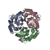
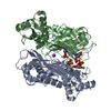
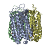
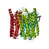
| Entry | Database: PDB / ID: 2jaf | ||||||
|---|---|---|---|---|---|---|---|
| Title | Ground state of halorhodopsin T203V | ||||||
 Components Components | Halorhodopsin | ||||||
 Keywords Keywords | MEMBRANE PROTEIN / CHROMOPHORE / CHLORIDE PUMP / ION TRANSPORT / MEMBRANE / CHLORIDE / RECEPTOR / ION PUMP / TRANSPORT / SENSORY TRANSDUCTION / PHOTORECEPTOR PROTEIN / TRANSMEMBRANE / RETINAL PROTEIN | ||||||
| Function / homology |  Function and homology information Function and homology informationphotoreceptor activity / phototransduction / monoatomic ion channel activity / plasma membrane Similarity search - Function | ||||||
| Biological species |  Halobacterium salinarum (Halophile) Halobacterium salinarum (Halophile) | ||||||
| Method |  X-RAY DIFFRACTION / SYNCHROTRON / MOLECULAR REPLACEMENT / Resolution: 1.7 Å X-RAY DIFFRACTION / SYNCHROTRON / MOLECULAR REPLACEMENT / Resolution: 1.7 Å | ||||||
 Authors Authors | Gmelin, W. / Zeth, K. / Efremov, R. / Heberle, J. / Tittor, J. / Oesterhelt, D. | ||||||
 Citation Citation | Journal: Photochem. Photobiol. / Year: 2007 Title: The crystal structure of the L1 intermediate of halorhodopsin at 1.9 angstroms resolution. Authors: Gmelin, W. / Zeth, K. / Efremov, R. / Heberle, J. / Tittor, J. / Oesterhelt, D. | ||||||
| History |
|
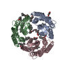
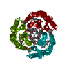
- Structure visualization
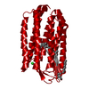
| Structure viewer | Molecule: MolmilJmol/JSmol |
|---|
- Downloads & links
-Download
| PDBx/mmCIF format | 2jaf.cif.gz | 62.8 KB | Display | PDBx/mmCIF format |
|---|---|---|---|---|
| PDB format | pdb2jaf.ent.gz | 44.9 KB | Display | PDB format |
| PDBx/mmJSON format | 2jaf.json.gz | Tree view | PDBx/mmJSON format | |
| Others |  Other downloads Other downloads |
-Validation report
| Arichive directory | https://data.pdbj.org/pub/pdb/validation_reports/ja/2jafftp://data.pdbj.org/pub/pdb/validation_reports/ja/2jaf | HTTPS FTP |
|---|
-Related structure data
| Related structure data |  2jagC 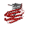 1e12S S: Starting model for refinement C: citing same article ( |
|---|---|
| Similar structure data |
-Links
 PDBj
PDBj


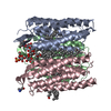
- Assembly
| Deposited unit | 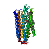
| ||||||||
|---|---|---|---|---|---|---|---|---|---|
| 1 | 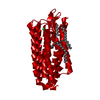
| ||||||||
| Unit cell |
|
-Components
-Protein / Sugars , 2 types, 2 molecules A
| #1: Protein | Mass: 28856.814 Da / Num. of mol.: 1 / Mutation: T203V Source method: isolated from a genetically manipulated source Source: (gene. exp.) Halobacterium salinarum (strain ATCC 29341 / DSM 671 / R1) (Halophile)Description: DSM 671 / Gene: hop, OE_1299R Production host: Halobacterium salinarum (strain ATCC 29341 / DSM 671 / R1) (Halophile)References: UniProt: B0R2U4 |
|---|---|
| #4: Sugar | ChemComp-BOG /  Type: D-saccharide / Mass: 292.369 Da / Num. of mol.: 1 Type: D-saccharide / Mass: 292.369 Da / Num. of mol.: 1Source method: isolated from a genetically manipulated source Formula: C14H28O6 / Comment: detergent*YM |
-Non-polymers , 4 types, 72 molecules 


| #2: Chemical |  Mass: 35.453 Da / Num. of mol.: 2 / Source method: obtained synthetically / Formula: Cl Mass: 35.453 Da / Num. of mol.: 2 / Source method: obtained synthetically / Formula: Cl#3: Chemical |  Mass: 256.424 Da / Num. of mol.: 2 / Source method: obtained synthetically / Formula: C16H32O2 Mass: 256.424 Da / Num. of mol.: 2 / Source method: obtained synthetically / Formula: C16H32O2#5: Chemical | ChemComp-RET / |  Mass: 284.436 Da / Num. of mol.: 1 / Source method: obtained synthetically / Formula: C20H28O Mass: 284.436 Da / Num. of mol.: 1 / Source method: obtained synthetically / Formula: C20H28O#6: Water | ChemComp-HOH / | Mass: 18.015 Da / Num. of mol.: 67 / Source method: isolated from a natural source / Formula: H2O |
|---|
-Details
| Compound details | ENGINEERED| Has protein modification | Y | |
|---|
-Experimental details
-Experiment
| Experiment | Method: X-RAY DIFFRACTION |
|---|
- Sample preparation
| Crystal | Density Matthews: 2.34 Å3/Da / Density % sol: 47.4 % |
|---|---|
| Crystal grow | Method: lipidic cubic phase Details: CRYSTALLIZED IN A CUBIC LIPID PHASE MADE OF 58-62 W/V % 1-MONOOLEIN, 3.3 M KCL, 3.5 MG/ML HR AND 25 MM TRIS/HCL, PH 7, R.T. |
-Data collection
| Diffraction | Mean temperature: 100 K |
|---|---|
| Diffraction source | Source: SYNCHROTRON / Site: ESRF  / Beamline: ID14-2 / Wavelength: 0.9393 / Beamline: ID14-2 / Wavelength: 0.9393 |
| Detector | Type: ADSC CCD / Detector: CCD |
| Radiation | Protocol: SINGLE WAVELENGTH / Monochromatic (M) / Laue (L): M / Scattering type: x-ray |
| Radiation wavelength | Wavelength: 0.9393 Å / Relative weight: 1 |
| Reflection | Resolution: 1.6→20 Å / Num. obs: 35303 / % possible obs: 95.9 % / Observed criterion σ(I): 3 / Redundancy: 8 % / Rmerge(I) obs: 0.14 / Net I/σ(I): 24.9 |
| Reflection shell | Redundancy: 7.9 % / Rmerge(I) obs: 0.74 / Mean I/σ(I) obs: 2.4 / % possible all: 89.5 |
- Processing
| Software |
| ||||||||||||||||||||||||||||||||||||||||||||||||||||||||||||
|---|---|---|---|---|---|---|---|---|---|---|---|---|---|---|---|---|---|---|---|---|---|---|---|---|---|---|---|---|---|---|---|---|---|---|---|---|---|---|---|---|---|---|---|---|---|---|---|---|---|---|---|---|---|---|---|---|---|---|---|---|---|
| Refinement | Method to determine structure: MOLECULAR REPLACEMENT Starting model: PDB ENTRY 1E12 Resolution: 1.7→20 Å / Data cutoff high absF: 10000 / Cross valid method: THROUGHOUT / σ(F): 2.4 Details: 3-10 HELIX DISTORTION BETWEEN LEU A 110 AND ALA A 113. PI-BULGE BETWEEN ALA A 178 AND TRP A 183 DISRUPTS HELIX E
| ||||||||||||||||||||||||||||||||||||||||||||||||||||||||||||
| Solvent computation | Bsol: 76.7206 Å2 / ksol: 0.388698 e/Å3 | ||||||||||||||||||||||||||||||||||||||||||||||||||||||||||||
| Displacement parameters |
| ||||||||||||||||||||||||||||||||||||||||||||||||||||||||||||
| Refinement step | Cycle: LAST / Resolution: 1.7→20 Å
| ||||||||||||||||||||||||||||||||||||||||||||||||||||||||||||
| Refine LS restraints |
|
