#1: Journal: Curr.Opin.Struct.Biol. / Year: 1998 Title: The Structure and Mechanism of the Family of Retinal Proteins from Halophilic Archaea Authors: Oesterhelt, D.
#2: Journal: J.Mol.Biol. / Year: 1995 Title: Three-Dimensional Structure of Halorhodopsin at 7 Angstrom Resolution Authors: Havelka, W. / Henderson, R. / Oesterhelt, D.
History
Deposition
Apr 14, 2000
Deposition site: PDBE / Processing site: PDBE
Revision 1.0
Jun 2, 2000
Provider: repository / Type: Initial release
Revision 1.1
Nov 14, 2012
Group: Atomic model / Database references ...Atomic model / Database references / Derived calculations / Non-polymer description / Other / Refinement description / Version format compliance
Revision 1.2
Feb 3, 2016
Group: Atomic model / Database references ...Atomic model / Database references / Derived calculations / Non-polymer description / Other / Refinement description / Source and taxonomy / Structure summary
Mass: 18.015 Da / Num. of mol.: 96 / Source method: isolated from a natural source / Formula: H2O
-
Details
Compound details
CHAIN A ENGINEERED MUTATION VAL229ALA
Has protein modification
Y
Sequence details
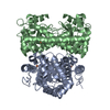
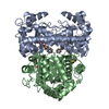
ALA A 229, MUTATION IN D2 STRAIN VERIFIED BY DIDEOXY-SEQUENCING C-TERMINUS NOT DEFINED IN ELECTRON ...ALA A 229, MUTATION IN D2 STRAIN VERIFIED BY DIDEOXY-SEQUENCING C-TERMINUS NOT DEFINED IN ELECTRON DENSITY FROM A263 - D274. IN THE CRYSTALS, HR ASSEMBLES SIMILARLY TO HOMOTRIMERS AS BACTERIORHODOPSIN.
-
Experimental details
-
Experiment
Experiment
Method: X-RAY DIFFRACTION / Number of used crystals: 1
-
Sample preparation
Crystal
Density Matthews: 2.54 Å3/Da / Density % sol: 41.5 %
Crystal grow
Method: lipidic cubic phase / pH: 7 Details: CRYSTALLIZED IN A CUBIC LIPID PHASE MADE OF 58-62 W/V % 1-MONOOLEIN, 4 M KCL, 3.3-4.0 MG/ML HR AND 50 MM TRIS/HCL, PH 7.
Resolution: 1.8→25 Å / Data cutoff high absF: 10000 / Isotropic thermal model: RESTRAINED / Cross valid method: THROUGHOUT / σ(F): 0 Details: LIPID PATCH BETWEEN HR TRIMERS MODELLED WITH 1-MONOOLEIN MOLECULES. NOTE THAT THESE OLC MOLECULES ARE MOSTLY ONLY PARTIALLY DEFINED BY ELECTRON DENSITY.
In the structure databanks used in Yorodumi, some data are registered as the other names, "COVID-19 virus" and "2019-nCoV". Here are the details of the virus and the list of structure data.
Jan 31, 2019. EMDB accession codes are about to change! (news from PDBe EMDB page)
EMDB accession codes are about to change! (news from PDBe EMDB page)
The allocation of 4 digits for EMDB accession codes will soon come to an end. Whilst these codes will remain in use, new EMDB accession codes will include an additional digit and will expand incrementally as the available range of codes is exhausted. The current 4-digit format prefixed with “EMD-” (i.e. EMD-XXXX) will advance to a 5-digit format (i.e. EMD-XXXXX), and so on. It is currently estimated that the 4-digit codes will be depleted around Spring 2019, at which point the 5-digit format will come into force.
The EM Navigator/Yorodumi systems omit the EMD- prefix.
Related info.:Q: What is EMD? / ID/Accession-code notation in Yorodumi/EM Navigator
Yorodumi is a browser for structure data from EMDB, PDB, SASBDB, etc.
This page is also the successor to EM Navigator detail page, and also detail information page/front-end page for Omokage search.
The word "yorodu" (or yorozu) is an old Japanese word meaning "ten thousand". "mi" (miru) is to see.
Related info.:EMDB / PDB / SASBDB / Comparison of 3 databanks / Yorodumi Search / Aug 31, 2016. New EM Navigator & Yorodumi / Yorodumi Papers / Jmol/JSmol / Function and homology information / Changes in new EM Navigator and Yorodumi
 Movie
Movie Controller


 Open data
 Basic information
 Components
 Keywords
 Function and homology information
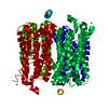
 HALOBACTERIUM SALINARUM (Halophile)
 X-RAY DIFFRACTION /
  Authors
 Citation
 Structure visualization
 Downloads & links
 Other downloads


 PDBj
PDBj


 Assembly
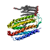


 Mass: 35.453 Da / Num. of mol.: 1 / Source method: obtained synthetically / Formula: Cl
 Mass: 39.098 Da / Num. of mol.: 1 / Source method: obtained synthetically / Formula: K
 Mass: 256.424 Da / Num. of mol.: 1 / Source method: obtained synthetically / Formula: C16H32O2
 Mass: 356.540 Da / Num. of mol.: 10 / Source method: obtained synthetically / Formula: C21H40O4
 Mass: 284.436 Da / Num. of mol.: 1 / Source method: obtained synthetically / Formula: C20H28O
 Sample preparation
 / Beamline: ID14-3 / Wavelength: 0.93
  Processing