Mass: 18.015 Da / Num. of mol.: 242 / Source method: isolated from a natural source / Formula: H2O
Sequence details
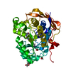
RESIDUES 20 TO 322. THIS SEQUENCE CORRESPONDS TO AN ISOFORM OF UNIPROT ENTRY Q15257: Q15257-2
-
Experimental details
-
Experiment
Experiment
Method: X-RAY DIFFRACTION / Number of used crystals: 1
-
Sample preparation
Crystal
Density Matthews: 2.16 Å3/Da / Density % sol: 39.5 %
Crystal grow
Temperature: 293 K / Method: vapor diffusion, hanging drop / pH: 7 Details: PTPA CRYSTALS WERE GROWN USING THE HANGING DROP METHOD AT 20C. 1UL OF PROTEIN (20 MG/ML) WAS MIXED WITH 1 UL OF PRECIPITANT (23% TO 28% PEG 4000, 0.2 M LITHIUM SULFATE, 0.1 M TRIS PH 7.0-8.5 ...Details: PTPA CRYSTALS WERE GROWN USING THE HANGING DROP METHOD AT 20C. 1UL OF PROTEIN (20 MG/ML) WAS MIXED WITH 1 UL OF PRECIPITANT (23% TO 28% PEG 4000, 0.2 M LITHIUM SULFATE, 0.1 M TRIS PH 7.0-8.5 AND 5 MM DTT) WERE EQUILIBRATED AGAINST 0.5 ML OF PRECIPITANT. LARGE PLATE CRYSTALS APPEARED IN 2 TO 3 DAYS. FOR CRYOPROTECTION 5% MPD WAS ADDED TO THE PRECIPITANT SOLUTION.
 Movie
Movie Controller


 Open data
 Basic information
 Components
 Keywords
 Function and homology information
 HOMO SAPIENS (human)
 X-RAY DIFFRACTION /
  Authors
 Citation
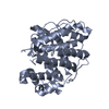
 Structure visualization
 Downloads & links
 Other downloads
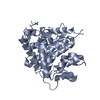


 PDBj
PDBj

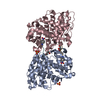
 Assembly
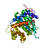

 Mass: 18.015 Da / Num. of mol.: 242 / Source method: isolated from a natural source / Formula: H2O
 Sample preparation
 / Beamline: ID14-1 / Wavelength: 0.9686
  Processing